
Scientific Imaging System

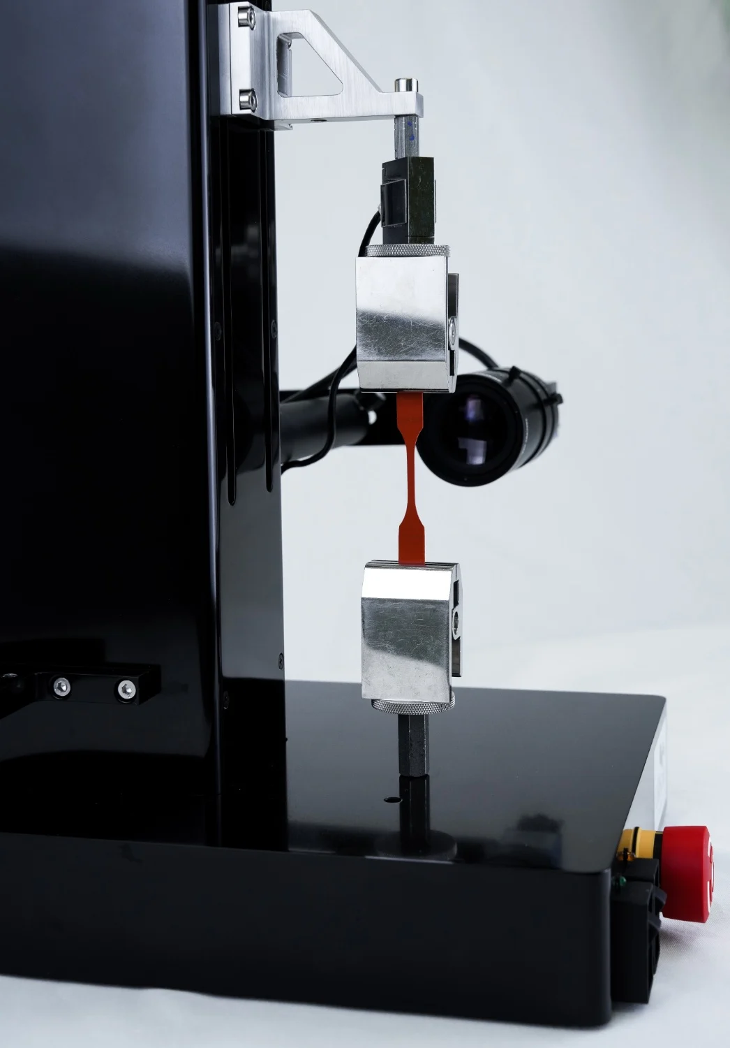
The Scientific Imaging System is a high performance camera and optics package that enables image-based strain measurement and strain mapping in CellScale’s LabJoy software. It supports real-time strain analysis for strain-controlled testing, and it is designed for image tracking workflows where you need clear, repeatable images across long tests or higher frequency capture routines.
Compatible with: UniVert, BioTester, MicroTester. The MicroTester G2 and BioTester 5000 & 6000 include this class of imaging system as standard.
When to choose the Scientific Imaging System: When you want image tracking for mechanical testing, including image-based strain measurement, strain mapping, and strain-controlled testing with improved imaging quality and repeatability for publications.
At a Glance
2048 × 2048 pixel imaging with USB 3.0 connectivity and up to 15 FPS full-frame capture
Enables strain mapping and image-based strain measurement for quantitative deformation tracking
Standard varifocal lens option (13 to 130 mm) for flexible field of view selection with manual adjustment
High-magnification option with microscope optics, 10x zoom, and a smaller field of view for fine features
Supports real-time strain analysis in LabJoy software for strain-controlled testing protocols
Why Use the Scientific Imaging System
A load cell alone cannot tell you whether deformation is uniform, whether grip slip is occurring, or whether strain localizes in the region you care about. The Scientific Imaging System adds an imaging pathway designed for quantitative tracking so LabJoy can compute image-based strain measurement and strain mapping, rather than relying on visual inspection or post hoc estimates.
Common reasons labs add the Scientific Imaging System:
- Run strain-controlled testing using image-derived strain instead of displacement assumptions
- Improve image tracking for mechanical testing when samples are small, low contrast, or prone to rotation
- Capture stable data for real-time strain analysis and repeatable reporting across users and templates
- Apply video extensometry style measurements when non-contact strain tracking is preferred
What’s Included

The Scientific Imaging System is supplied as a camera and optics package integrated for LabJoy workflows.
Package options typically include:
- High resolution scientific camera with USB 3.0 connectivity
- Standard varifocal lens option (13 to 130 mm) with manual adjustment
- Optional high-magnification microscope optics (10x zoom) for small fields of view
- Mounting and integration components appropriate to the instrument configuration
Typical Workflows
The Scientific Imaging System is used when your protocol depends on quantifying deformation optically. In LabJoy, image streams support image-based strain measurement and strain mapping, enabling real-time strain analysis during the test.
Digital Image Correlation (DIC)
Image-based tracking configurations in LabJoy for non-contact deformation measurement, including region-of-interest strain mapping
With image-based strain measurement to track elongation, detect grip slip, and validate alignment during loading
With image tracking to quantify time-dependent strain under load, especially when displacement is not representative of specimen strain
Where strain mapping helps quantify non-uniform deformation in soft or heterogeneous specimens
Using a fixed deformation and optical strain tracking to improve confidence in relaxation curves
Hydrated and Temperature Controlled Testing
Mapping runs in a bath when hydration and temperature stability matter across a long sequence
Instrument Compatibility
Works with:
- UniVert S and UniVert 1kN: upgrade from the standard webcam imaging setup
- BioTester 3000: optional imaging upgrade
- BioTester 5000/6000: Included with the instrument
- MicroTester G2: Included with the instrument; add an optional secondary camera for side view (great for shear testing)
Important note: MicroTester LT uses a different imaging configuration and cannot be upgraded to the Scientific Imaging System.

Specifications
| Resolution | 2048 × 2048 pixels |
| Connectivity | USB 3.0 |
| Full-frame capture | Up to 15 FPS, with higher speeds for regions of interest |
| Standard varifocal lens | 13 to 130 mm (manual adjustment), with an approximate 5 to 50 mm field of view |
| High-magnification option | microscope optics with 10x zoom and an approximate 1 to 6.5 mm field of view |
Research Applications Supported
The Scientific Imaging System is most useful when deformation tracking and strain calculation are central to the experiment, including image tracking for mechanical testing, strain mapping, and video extensometry style measurements.
Heart Valve Tissue Engineering & Mechanics
Cartilage & Meniscus Mechanics
Polymers & Elastomers Testing
Membranes & Thin Films Mechanics
Scaffold Mechanical Testing
Lung & Pleural Tissue Biomechanics
Material Fatigue & Durability
Wearable Bioelectronics
FAQs
What does the Scientific Imaging System add compared to standard imaging?
The high resolution scientific camera is designed for quantitative tracking so LabJoy can run image-based strain measurement and strain mapping, rather than using basic video capture only. It also supports real-time strain analysis for protocols that depend on image-derived strain.
Can I use the Scientific Imaging System for strain-controlled testing?
Yes. The Scientific Imaging System supports strain-controlled testing by enabling image-derived strain signals, which is useful when specimen deformation is not well represented by actuator displacement.
Is this the same camera used on the MicroTester G2 and BioTester systems?
Mostly. The camera body is the same, but the lens, mounting, and zoom capabilities differ by system. On the MicroTester G2, a second viewing axis can be added for a side view, using the same camera type (this is particularly helpful with the shear axis on the MicroTester G2).
Is video extensometry supported?
Yes. Many users apply video extensometry style workflows using LabJoy image tracking for mechanical testing, especially when contact extensometers are impractical or when non-contact strain measurement is preferred. Video extensometry can also complement strain mapping when strain localizes in a specific region.
Can I upgrade a MicroTester LT to the Scientific Imaging System?
No. MicroTester LT uses a different camera configuration and cannot be upgraded to the Scientific Imaging System.