


In our new In the Lab blog series, we catch up with researchers using CellScale systems.
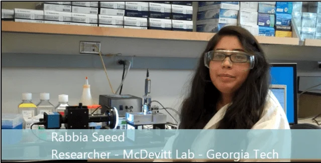
In our first blog, Caleb Horst chats with Rabbia Saeed of the McDevitt Lab at Georgia Tech (Dr. McDevitt is now a Senior Investigator at the Gladstone Institute of Cardiovascular Disease at UC San Francisco).
Quantifying the Mechanics of Cell Spheroids at the Microscale
Researchers at Georgia Tech are investigating how the mechanical properties of embryonic stem cell spheroids influence cell differentiation and tissue organization. Using the CellScale MicroSquisher (now the MicroTester), the team performs microscale compression, stress relaxation, and creep testing on three-dimensional cell aggregates that fall between the size limits of atomic force microscopy and conventional mechanical testers.
This work highlights how direct mechanical testing of spheroids enables quantitative insights into viscoelastic behavior that cannot be inferred from morphology alone.
Why Cell Spheroid Mechanics Matter
Cell spheroids occupy a unique experimental space. They are larger and more structurally complex than single cells, yet far smaller and softer than bulk tissues. Because of this, traditional mechanical testing approaches struggle to characterize them accurately.
Understanding spheroid mechanics is increasingly important for:
Stem cell differentiation studies
Cancer and tumor modeling
Drug screening in 3D culture systems
Tissue self-organization and morphogenesis research
Mechanical stiffness, stress relaxation, and creep behavior all provide insight into how cells interact with and remodel their surrounding microenvironment.
Why the MicroSquisher Is Ideal for Micromass Aggregates
Caleb Horst: Rabbia, I understand that you are researching the mechanical properties of embryonic stem cell spheroids to determine the relationship between stiffness and cell differentiation. Can you tell me a bit about why you chose CellScale’s Microsquisher for your testing?
Rabbia Saeed: The McDevitt Lab has chosen the MicroSquisher for our testing because our samples are not tissues but they’re not single cells either. Rather, our samples are micromass aggregates of stem cells. So due to the size and the structure of our aggregates, we can’t use Instron machines or atomic force microscopy to get the mechanical properties of our samples. However, the MicroSquisher does have the perfect 50 micron to 20 millimeter scale needed to test our samples.
CH: I know a lot of researchers do different things with the MicroSquisher, so I was hoping you could walk us through a typical test setup for your application.
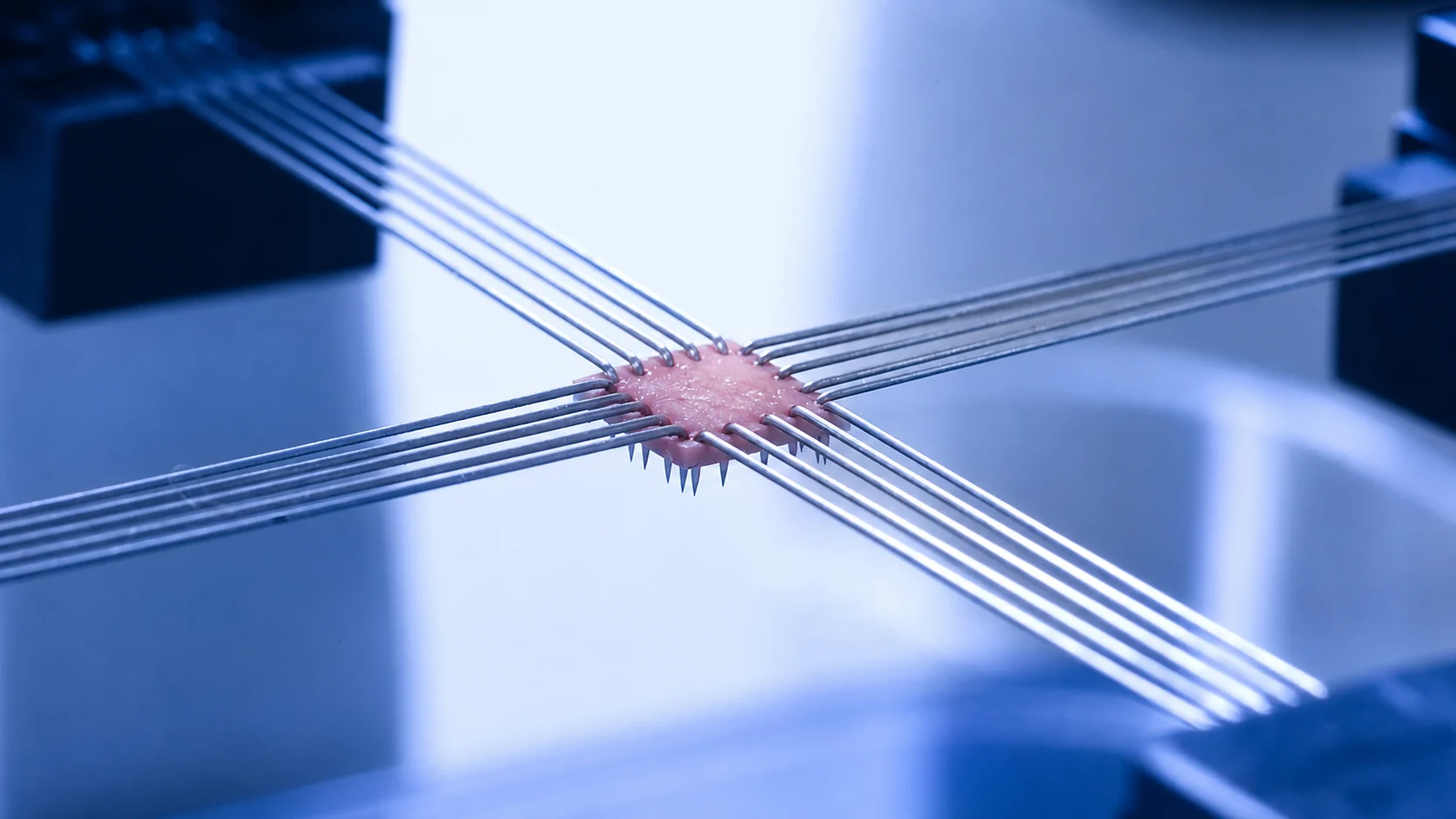
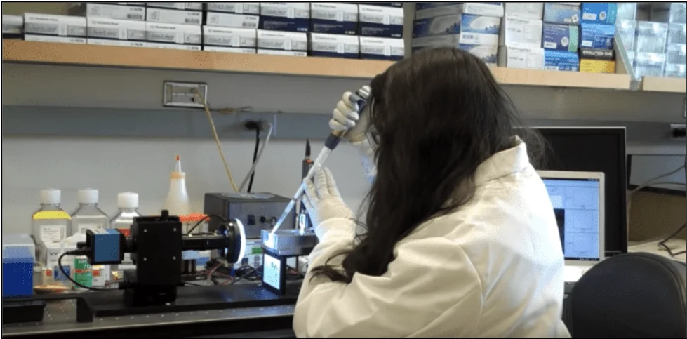
RS: The procedure for setting up the MicroSquisher for our testing is very simple. Because the MicroSquisher relies on cantilever mechanics, the first step is to load a cantilever beam into the grip. The grip is then powered by 3 piezoelectric motors that go in the X, Y, and Z directions to orient the cantilever. Then our next step is that we use a pipette to transfer one of our samples into the fluid bath [Figure 1]. After this, we can then orient the cantilever over the sample with the piezoelectric motors and we’re ready to squish.
Compression, Stress Relaxation, and Creep Testing of Spheroids
One of the key advantages of the MicroSquisher is its ability to perform multiple mechanical test modes on intact spheroids.
CH: Rabbia, can you tell me a little bit more about the test results you’re getting, and why you find them interesting?
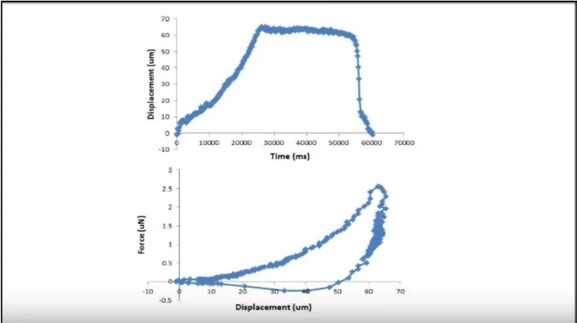
RS: Absolutely. One of the tests that we acquire is the force-displacement curve [Figure 2] or the stress-strain curve, if you normalize the area.
So, essentially what the MicroSquisher does is it has a ramp function where you can compress the sample, hold the sample, and then decompress the sample, which is called the recovery phase. And then this sort of test produces a typical stress-strain curve, where you can observe the hysteresis of our viscoelastic samples.
CH: As well as doing force-displacement tests, I understand you’re also doing some stress relaxation work?
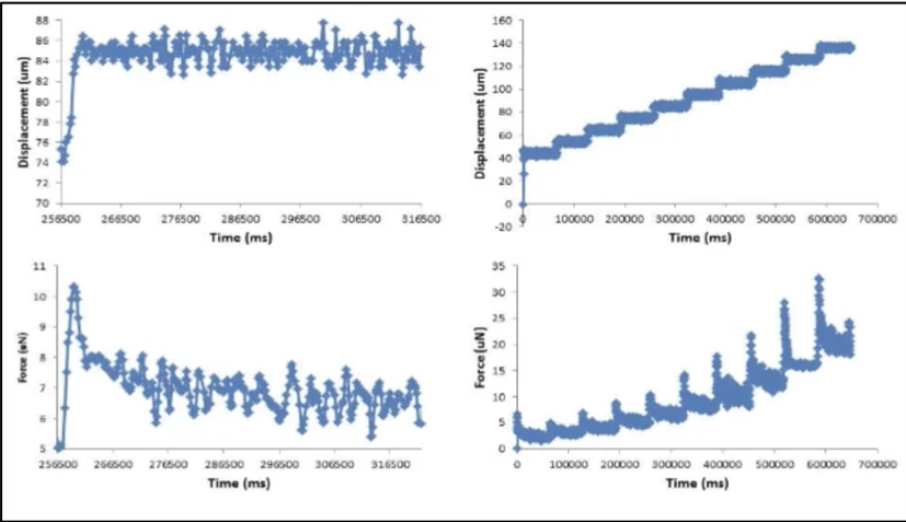
RS: Absolutely. We have been acquiring a variety of stress-relaxation curves. The MicroSquisher applies a unit step at a constant deformation. Here you can see it’s approximately 86-85 micronewtons and the resulting relaxation of our samples is seen below [Figure 3]. There’s a peak force and then equilibrium force as our samples relax.
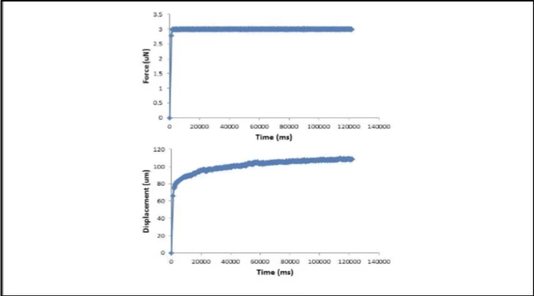
The last data type that the MicroSquisher can acquire is called the creep curves. So, we apply a constant force – as you can see, this perfectly applied unit step at 3 microNewtons. We then measure the change in deformation at that constant force [Figure 4].
Linking Spheroid Mechanics to Functional Cell Behavior
CH: So, I understand you’ve been using the MicroSquisher for some time now. Have you been successful in collecting data that has enabled you to learn new and interesting things? And have you started to put together data that’s useful in publications?
RS: The McDevitt Lab has had the MicroSquisher for about a year and a half now, and in that time we already have a publication by Dr. Priya Baraniak that demonstrates how microparticle incorporation stiffened spheroids of mesenchymal stem cells. With the help of the MicroSquisher, we will certainly have at least one more publication on the mechanical properties of stem cell aggregates in the near future, so stay on the lookout for another McDevitt Lab publication!
CH: Thanks for your time Rabbia. I’m looking forward to seeing more amazing research coming out of McDevitt Lab.
Watch the interview online: https://www.youtube.com/watch?v=f26rz37ko28
Why This Work Still Matters
This early work at Georgia Tech helped establish microscale mechanical testing as a practical and necessary tool for studying three-dimensional cell models. Today, similar approaches are widely used in:
Tumor spheroid mechanics
Organoid stiffness matching
Drug response screening in 3D cultures
Tumor microenvironment research
By treating stiffness as a measurable physical property, rather than an inferred characteristic, researchers can more accurately connect molecular changes to functional outcomes.
Learn More
Product: CellScale MicroTester (upgraded form of the MicroSquisher)
Related Applications: Cancer Mechanobiology, Organoid & Tissue Mimetic Systems
Related Research: Tumor stiffness and CAF-driven matrix remodeling