A hydrogel can look perfectly fine after bioprinting, but that doesn’t necessarily tell you much about how the cells inside it will behave. Two constructs made from the same material might hold their shape equally well and still have different mechanical properties. Depending on what the researchers are studying, those differences can matter quite a bit.
A recent study from the University of California San Diego looked at this question using human neural progenitor cells encapsulated in gelatin methacrylate (GelMA). The researchers were developing a 3D neural tissue model for studying the effects of environmental pollutants, particularly chlorpyrifos, a pesticide known to affect the nervous system. As part of the work, they wanted to see whether cells grown in softer and stiffer hydrogels would respond differently to the same chemical exposure.
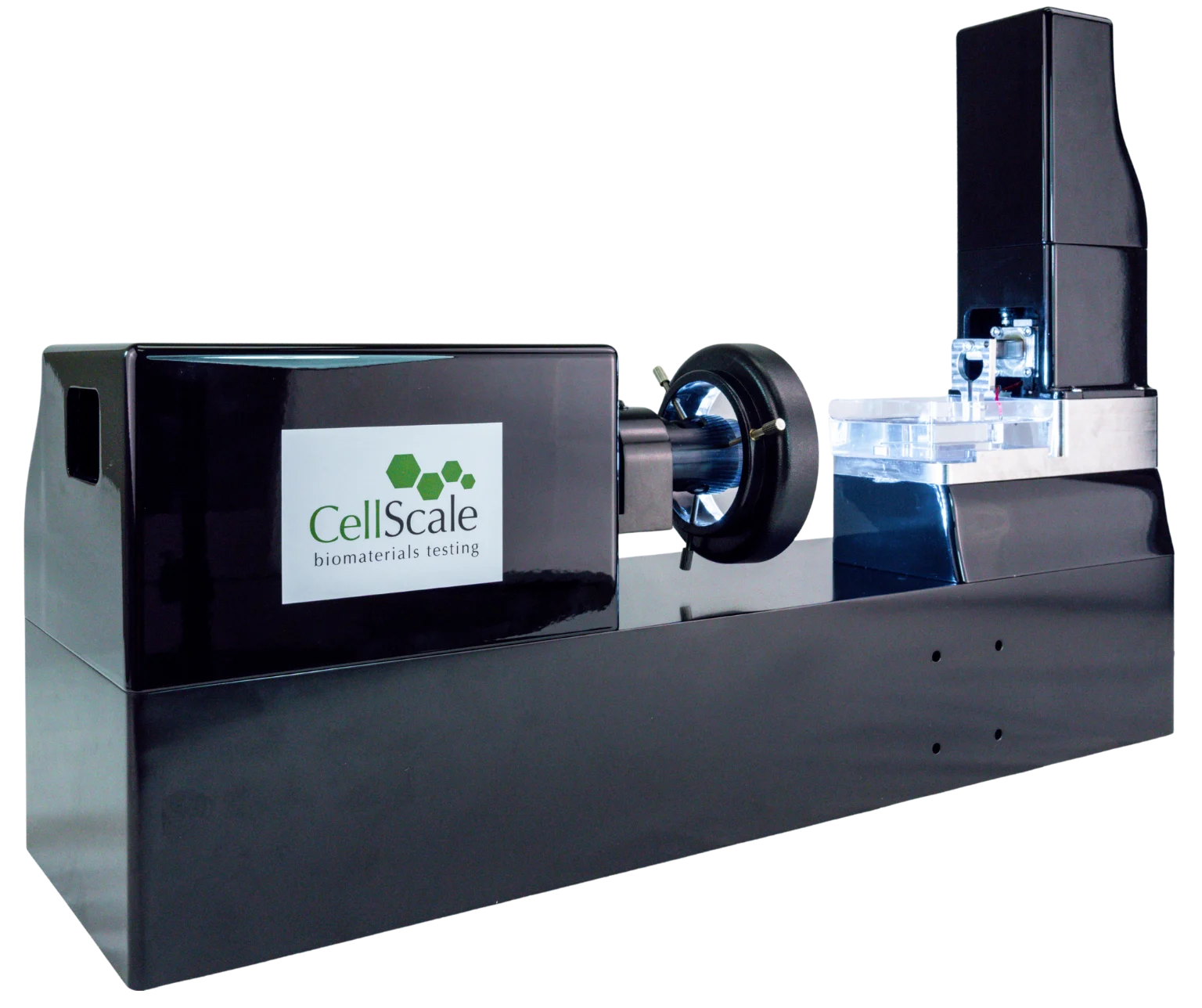
They used digital light processing (DLP) bioprinting to make the constructs, adjusting the light exposure to change the properties of the GelMA. The resulting hydrogels were then mechanically characterized using a CellScale MicroTester, which measured compressive Young’s moduli of approximately 2 kPa and 4 kPa.
What followed was an interesting comparison. The two groups contained the same type of neural progenitor cells and the same nominal GelMA concentration, but their responses to pollutant exposure were not quite the same. Cell viability differed, as did the expression of several genes associated with neural progenitor cell activity and responses to injury.
The work, published by Yao et al. in the International Journal of Bioprinting, provides a useful example of why GelMA hydrogel stiffness is worth measuring when mechanical properties are being used as a variable in a biological experiment.
Why GelMA Hydrogel Stiffness Matters in Neural Tissue Engineering
Neural cells are sensitive to their surroundings. In the brain, they’re packed into a three-dimensional environment where they interact with neighboring cells and the extracellular matrix. That environment is mechanically soft, and its properties can affect how cells grow, move, and develop.
It’s a difficult environment to reproduce in the lab. Conventional two-dimensional cultures are useful for many experiments, but cells grown on a flat surface don’t experience quite the same physical conditions as cells surrounded by a three-dimensional matrix.
Hydrogels offer another approach. They contain large amounts of water, can support embedded cells, and can be made with mechanical properties that more closely resemble soft biological tissues.
GelMA is frequently used for this purpose. It is derived from gelatin and contains methacrylate groups that allow it to be crosslinked using light. Researchers can alter the formulation or photocrosslinking conditions to produce materials with different stiffnesses.
That flexibility is useful, although it also raises a question: when two groups of cells are grown in GelMA hydrogels with different mechanical properties, how much does the surrounding matrix affect what the cells do?
It’s something researchers working in neural tissue mechanics have been investigating, particularly as 3D culture methods become more common.
In the UC San Diego study, the researchers weren’t trying to determine the best possible stiffness for neural tissue. They were interested in how human neural progenitor cells responded to toxic chemicals, and whether that response differed between two mechanical environments.
To make that comparison, they first needed a way to produce soft and stiff constructs that could support the cells. Then they needed to find out how stiff those constructs actually were.
Producing Two GelMA Hydrogels with Different Stiffnesses
The team started with a bioink containing 10% (w/v) GelMA and 0.6% (w/v) lithium phenyl-2,4,6-trimethylbenzoylphosphinate (LAP), a photoinitiator. Human induced pluripotent stem cell-derived neural progenitor cells were mixed into the material before printing.
They used an in-house DLP bioprinter to produce small, brain-shaped constructs. Rather than depositing material through a nozzle, the system projected patterned light onto the bioink to crosslink selected regions.
For the softer group, the researchers used 365 nm light at 6.8 mW/cm² for 20 seconds. For the stiffer group, the exposure was increased to 10.8 mW/cm² for 30 seconds.
The exposure doses were 136 and 324 mJ/cm², respectively.
Nothing about the nominal GelMA concentration changed between those two conditions. The researchers were instead using the printing process itself to alter the finished material.
This is one of the practical considerations when working with 3D bioprinting and bioink materials. The formulation is only part of what determines the properties of a printed scaffold. Light exposure, crosslinking conditions, and other processing parameters can change the mechanical behavior of the final construct.
The researchers also had to consider cell density. Early experiments used 15 million cells/mL, but they later increased this to 50 million cells/mL after observing better cell viability at the higher density during the initial exposure period.
Once printed, the cell-laden hydrogels were maintained in culture. The researchers monitored the cells over seven days, using microscopy and a luminescence-based assay to follow their growth.

The cells remained distributed throughout the printed structures, and the luminescence measurements increased considerably between days three and seven. Some of the microscopy images also showed cellular extensions developing within the constructs.
The hydrogels were therefore supporting cell growth under the conditions examined. But the two exposure settings were intended to produce different mechanical environments, and that still needed to be checked experimentally.
Measuring GelMA Hydrogel Stiffness with the MicroTester
To measure the stiffness of the bioprinted samples, the researchers used CellScale’s MicroTester.
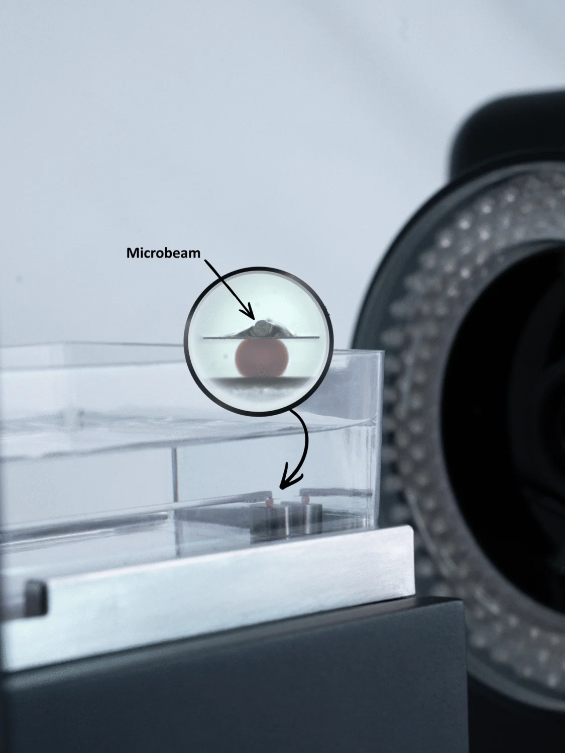
These were small, compliant hydrogel constructs, rather than the larger cylindrical specimens often used in conventional material testing. Their size is one reason micro-mechanical testing was appropriate for this part of the work.
The authors report that each sample underwent three compression cycles using a 0.3048 mm diameter stainless steel beam and platens. They recorded force and displacement during loading, then processed the measurements using custom MATLAB scripts to calculate the compressive modulus.
How Was Young's Modulus Measured in the Bioprinted GelMA Hydrogels?
Young’s modulus is commonly used to describe how much a material resists deformation under an applied load. In compression testing, force and displacement measurements can be converted into stress and strain using the specimen geometry. The relationship between stress and strain is then used to estimate the modulus.
For hydrogels, the calculation isn’t always as straightforward as it might seem. The result can depend on the amount of compression, the loading rate, the specimen dimensions, and the region of the stress-strain curve used for analysis.
The publication doesn’t provide every detail of the modulus calculation. It describes the three compression cycles and the use of MATLAB to process the force-displacement data, but doesn’t specify the loading rate, maximum deformation, or fitting interval. Those details would be needed to reproduce the test exactly.
The reported results were nevertheless quite different between the two groups.
The soft GelMA constructs had a compressive Young’s modulus of approximately 2 kPa, while the stiff constructs measured approximately 4 kPa.
So, with the same nominal GelMA formulation, the higher-intensity and longer light exposure produced a hydrogel with roughly twice the measured modulus.
The measurements were useful because they established what the printing changes had done to the finished material. Without them, the researchers would have known only that one group received more light exposure than the other.
This is a recurring issue in hydrogel mechanical testing. A change in formulation or fabrication conditions doesn’t always translate into a predictable change in modulus, particularly when working with highly hydrated materials.
In this case, the researchers had two mechanically distinct groups they could carry forward into the biological comparisons.
Did GelMA Hydrogel Stiffness Affect the Response to Chlorpyrifos?
Chlorpyrifos is an organophosphate pesticide that has been studied extensively for its effects on the nervous system. One of its known toxic mechanisms involves acetylcholinesterase, the enzyme responsible for breaking down the neurotransmitter acetylcholine.
The researchers exposed their bioprinted neural tissue models to chlorpyrifos and measured cell viability after three and five days. They also examined gene expression to look for differences in how the cells were responding.
One thing that stands out is the difference in viability between the soft and stiff constructs.
After three days of exposure, the stiffer group had a mean viability of roughly 0.55, compared with approximately 0.40 in the softer group. Viability was considerably lower in both groups after five days, although the stiff constructs still showed a higher measured value.
The authors also examined three genes associated with neural progenitor cell biology: PAX6, Nestin, and SOX2.
PAX6 and Nestin expression were higher in the stiffer constructs, whereas SOX2 expression was higher in the softer ones. These markers are involved in different aspects of neural progenitor cell identity, development, and responses to injury.
The researchers then looked at several Alzheimer’s disease-associated markers, including beta amyloid, BACE1, and AChE. The softer group generally showed higher expression of these markers in the reported comparison.
These are transcriptional measurements, so they shouldn’t be interpreted as direct evidence of protein accumulation, enzyme activity, or Alzheimer’s disease development. They do, however, show differences in cellular response between the two experimental conditions.

The authors discuss these findings in relation to how matrix mechanics may affect the response of neural progenitor cells to injury. Higher Nestin expression in the stiff group, for example, may reflect a different cellular stress or remodeling response, while the higher SOX2 expression in the soft group may be related to the maintenance of progenitor cell identity.
There is some uncertainty in what can be attributed specifically to stiffness. The two groups were produced using different photocrosslinking conditions, which may have changed other aspects of the GelMA network as well. And gene expression provides only one view of what the cells are doing.
Even so, the comparison is worth examining. A twofold difference in measured compressive modulus was accompanied by changes in cell viability and several markers associated with neural progenitor cell behavior.
For researchers studying stem cell mechanobiology, this is one reason to pay attention to the mechanical properties of the matrix, even when the main experiment is focused on a chemical or biological treatment.
Looking More Closely at Cell Viability in the Printed Constructs
Before comparing the soft and stiff groups, the researchers spent some time adjusting the conditions used in their neural tissue model.
They first tested chlorpyrifos concentrations of 5, 20, and 500 µg/L. At the highest concentration, cell viability was substantially lower than at the two lower concentrations. This provided a measurable toxic response that could be examined in subsequent experiments.
They also compared two cell densities, 15 million and 50 million cells/mL.
The higher-density constructs had better viability after three days of chlorpyrifos exposure. By day five, however, both groups showed very low viability, and the difference between them was no longer statistically significant.
The microscopy images give a more direct view of what was happening. Cells were stained using fluorescent markers, with live cells appearing green and dead cells red.

At day zero, the fluorescence images show a strong green signal throughout the constructs. By day three, there are fewer visibly live cells and more red staining. By day five, the images show a further shift toward dead-cell staining.
This part of the study isn’t directly about hydrogel stiffness, but it helps explain how the researchers arrived at their final experimental conditions.
It also shows that the viability measurements were influenced by more than the mechanical environment. Cell density, pollutant concentration, and exposure duration all affected the results.
Testing Another Pollutant in the Bioprinted Neural Tissue Model
After investigating chlorpyrifos, the researchers examined its metabolite, chlorpyrifos-oxon, as well as another environmental pollutant, benzo[a]pyrene (BaP).
Chlorpyrifos-oxon is formed when chlorpyrifos is metabolized in the body. Because the bioprinted model didn’t contain a liver or other system for metabolizing the pesticide, the researchers added the metabolite directly to the neural tissues.
The two compounds produced different patterns of toxicity. Chlorpyrifos caused a larger overall decrease in cell viability, while chlorpyrifos-oxon produced stronger changes in some of the measured gene-expression markers, including Nestin and beta amyloid.
The team then tested BaP, a compound found in sources such as vehicle exhaust, cigarette smoke, and other combustion products.
Once again, the researchers used both the soft and stiff GelMA constructs.
Viability was already very low in both groups after three days of BaP exposure. At day five, the stiffer constructs showed higher measured viability than the softer ones, although both remained at low levels. The researchers also reported increased SOX2 expression and decreased AChE expression in the BaP-treated cells.

The authors see this as an early version of a more complex tissue model. They note that additional cell types, particularly astrocytes, would be useful for examining interactions involved in neural injury. A blood-brain barrier and dynamic culture conditions could also be incorporated in later versions.
For now, the model provides a way to investigate pollutant responses in human neural progenitor cells grown within a defined 3D hydrogel environment.
Why Mechanical Testing Matters for 3D Bioprinted Tissue Models
One of the difficulties with 3D bioprinting is that the same nominal bioink formulation doesn’t always produce the same mechanical properties. Printing parameters matter, and relatively small changes in processing can affect the material that the cells ultimately experience.
In the UC San Diego study, the team kept the GelMA concentration constant but varied the light exposure. MicroTester compression measurements confirmed that the finished constructs had different moduli.
Those measurements gave the researchers a way to describe the mechanical conditions used in the subsequent toxicity experiments. The biological results could then be compared alongside measured material properties rather than printing settings alone.
There are similar considerations in other tissue engineering applications. Previous research on GelMA bioink mechanics in 3D bioprinting looked at how changes in bioink composition affected mechanical behavior. Another study, examining tunable scaffold stiffness for in vitro liver models, provides a different example of stiffness being considered in the design of an engineered tissue environment.
The way stiffness is measured also deserves some attention. Compression, indentation, tension, and rheology can produce different results, even when applied to nominally similar hydrogels. The specimen geometry, hydration conditions, loading rate, and material response all play a role.
Our earlier article on hydrogel stiffness measurement discusses these differences across several mechanical testing methods.
For very small printed constructs, the testing setup introduces additional considerations. The specimen needs to be positioned carefully, the applied forces may be low, and deformation can be difficult to measure accurately.
In this study, the reported 2 kPa and 4 kPa values were enough to establish two distinct mechanical conditions. The researchers could then examine how cells behaved in those environments, while recognizing that additional work would be needed to separate the effects of stiffness from other changes associated with photocrosslinking.
Using the MicroTester for Small Hydrogels and Engineered Tissues
The CellScale MicroTester is designed for mechanical testing of small biological tissues, hydrogels, and other soft materials where the forces involved can be too low for conventional mechanical testing equipment.
Depending on the specimen, researchers may perform compression, indentation, tension, or other microscale tests while recording the applied force and displacement.
For a soft hydrogel, compression measurements can be used to calculate a modulus when the sample geometry and analysis method are appropriate. Other experiments may focus on how force changes with displacement, how a small tissue responds to repeated loading, or how mechanical behavior differs between groups of specimens.
The UC San Diego researchers used the MicroTester for a fairly specific task: measuring the compressive modulus of small GelMA neural tissue constructs produced under two different light exposure conditions.
The results showed that the softer specimens had a Young’s modulus of approximately 2 kPa, compared with around 4 kPa for the stiffer group.
That information became part of the experimental comparison rather than simply a material property reported on its own. The researchers went on to examine cell viability and gene expression following pollutant exposure, with the measured stiffness providing context for the different cellular responses.
For researchers developing 3D tissue models, this is one way mechanical testing can fit into the workflow. Once a printed or fabricated construct has been mechanically characterized, its properties can be considered alongside cell culture, imaging, and biochemical measurements when evaluating the model.