Measuring Tissue Surface Tension in Zebrafish Embryo Aggregates | IST Austria

Research Highlights & Case Studies
Quick Overview:
Micro-scale compression testing of zebrafish embryo cell aggregates to quantify tissue surface tension and mechanical changes during early development.

Researchers at the Institute of Science and Technology Austria (IST Austria) study how mechanical properties guide cell sorting, aggregation, and tissue organization during early embryonic development.

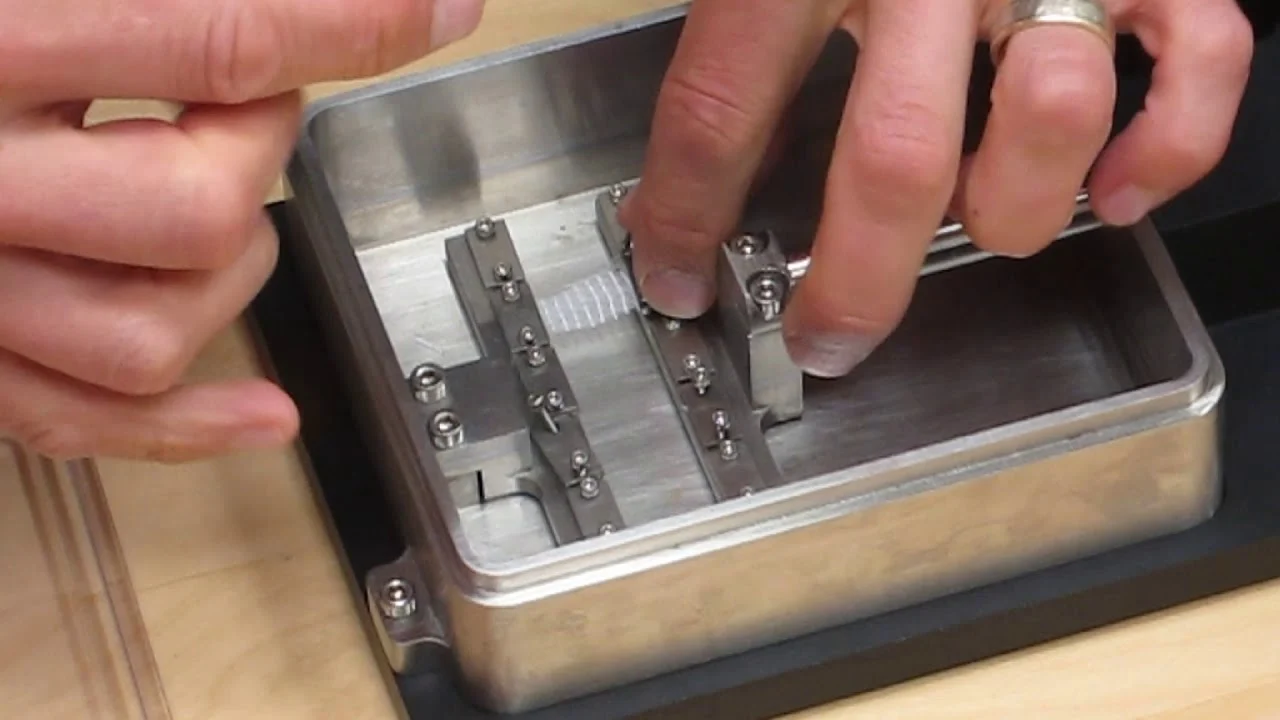
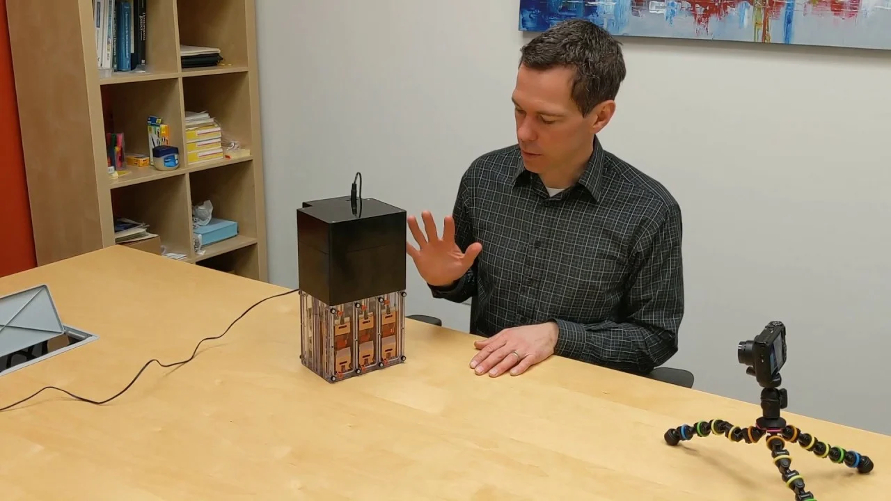
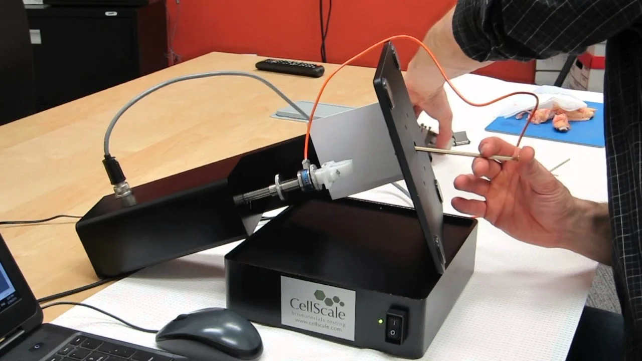
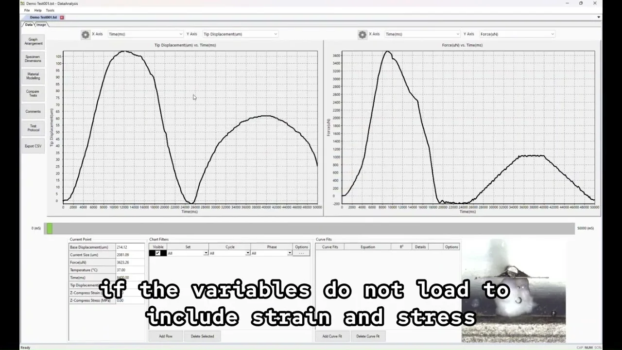
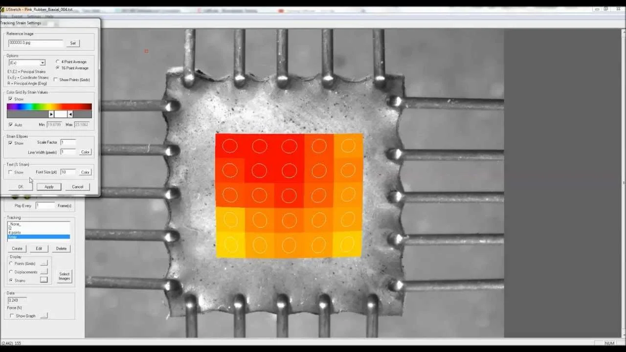
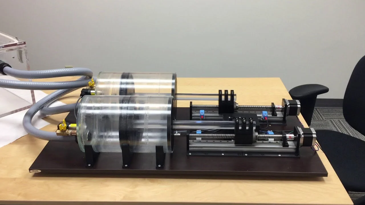
In this interview, Gabby Krens from the lab of Prof. Carl-Philipp Heisenberg explains how micro-scale mechanical testing is used to measure the surface tension and viscoelastic behavior of zebrafish embryo cell aggregates. By compressing spherical tissue aggregates and measuring force relaxation, the team quantifies physical properties that influence tissue rearrangement and differentiation over time.

This work highlights how mechanical measurements complement imaging and molecular biology approaches in developmental biology and mechanobiology research.

Related Videos:

MechanoCulture T6 Uniaxial Mechanical Stimulation Demonstration

MechanoCulture J1 Mechanical Stimulation System Introduction

Biomaterials Mechanical Testing for the Classroom Using the UniVert

Stress Strain Curve Data Analysis

Mechanical Testing Data Review and Analysis Software Demonstration

Mechanical Test Data Analysis and Strain Measurement Software Overview

Real-Time Image-Based Strain Control for Mechanical Testing

Biaxial Image-Based Strain Tracking

Image Tracking and Strain Analysis Software Demonstration

Custom Pressure–Volume Control System for Lung Mechanics Testing

Contact Sales

Product of Interest: