Imaging Collagen Fiber Orientation During Biaxial Heart Valve Testing

Research Highlights & Case Studies
Quick Overview:
Polarization-based imaging of collagen fiber orientation during biaxial heart valve testing.

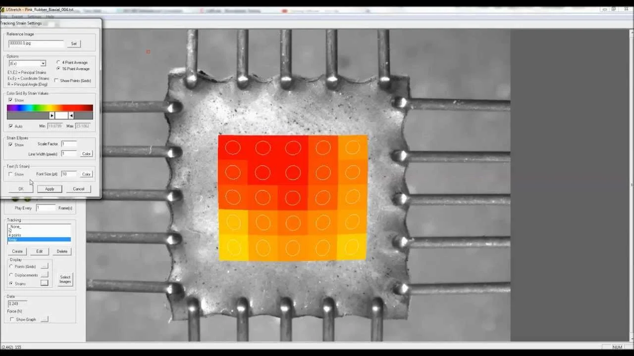
This research presentation showcases the development of a polarization-based imaging system for quantifying collagen fiber orientation in heart valve tissue during biaxial mechanical testing.

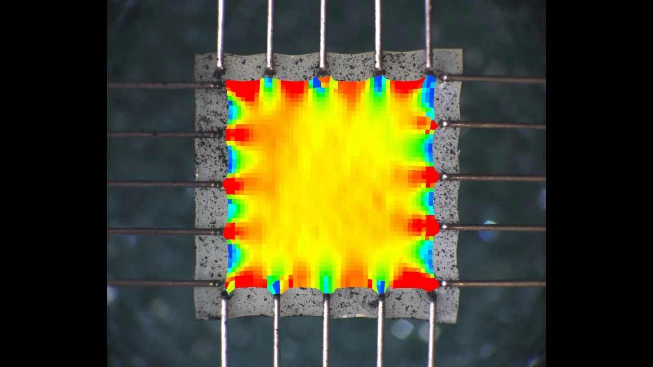
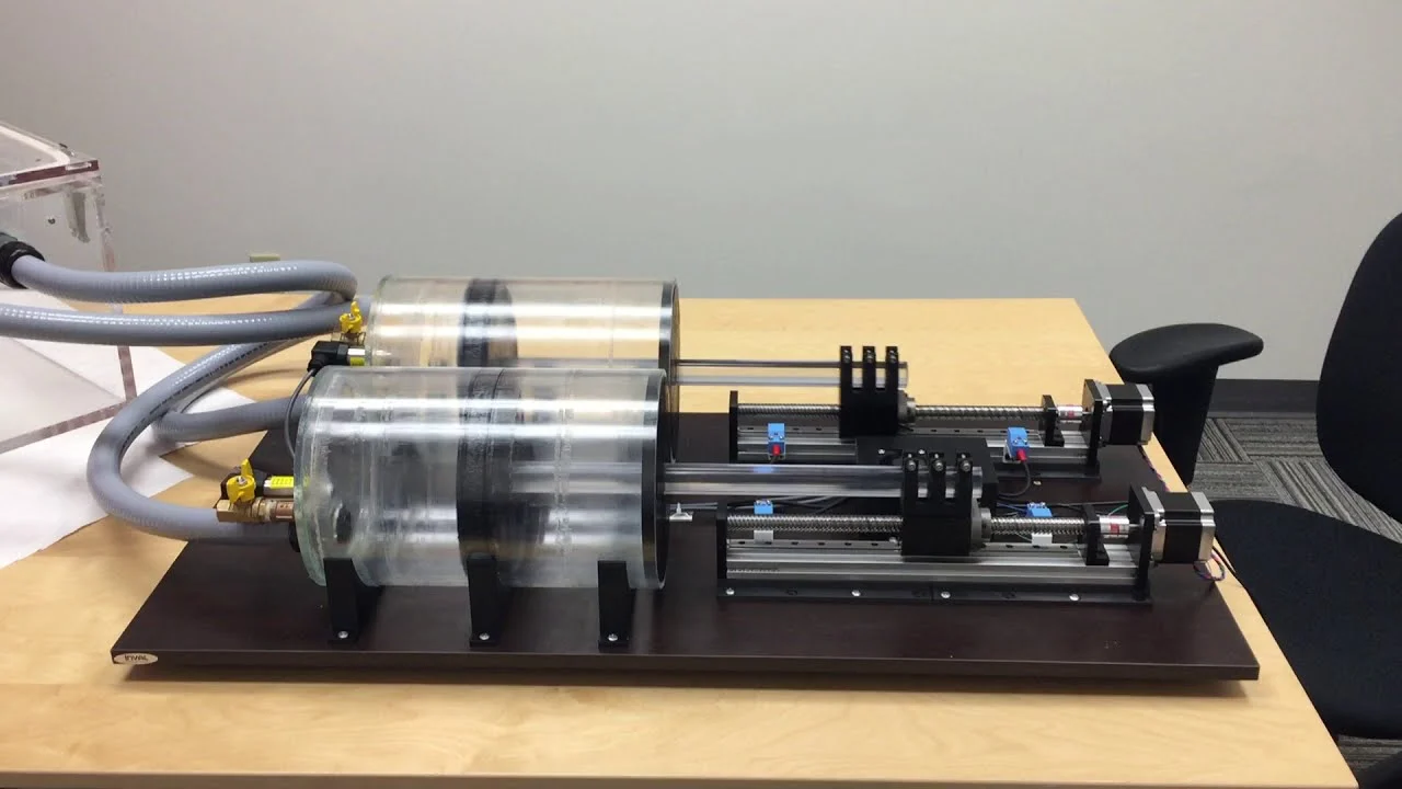
Sponsored by Prof. Chung-Hao Lee at the University of Oklahoma, this capstone project demonstrates how rotating polarizers and polarization-dependent light reflection can be used to dynamically map collagen fiber orientation while tissue is mechanically loaded.

When paired with a biaxial testing system, this modular imaging approach enables real-time visualization of microstructural changes in fibrous soft tissues, supporting advanced research in heart valve biomechanics and constitutive modeling.

Related Videos:

MechanoCulture T6 Uniaxial Mechanical Stimulation Demonstration

MechanoCulture J1 Mechanical Stimulation System Introduction

Biomaterials Mechanical Testing for the Classroom Using the UniVert

Stress Strain Curve Data Analysis

Mechanical Testing Data Review and Analysis Software Demonstration

Mechanical Test Data Analysis and Strain Measurement Software Overview

Real-Time Image-Based Strain Control for Mechanical Testing

Biaxial Image-Based Strain Tracking

Image Tracking and Strain Analysis Software Demonstration

Custom Pressure–Volume Control System for Lung Mechanics Testing

Contact Sales

Product of Interest: