


Vascular bioprinting is gaining attention because it offers a way to fabricate tissue engineered vascular grafts with more control over geometry, composition, and cell distribution than many traditional scaffold fabrication methods. This matters because small-diameter vascular grafts remain a major challenge in cardiovascular repair. Even though autologous vessels are still preferred, graft failure can still result from poor patency and compliance mismatch. That is why customized tissue engineered vascular grafts continue to be an important goal in vascular tissue engineering.
In this study, researchers from Binghamton University and Syracuse University developed a new approach for vascular bioprinting using a rotary 3D bioprinter and a gelatin-fibrinogen bioink. CellScale contributed to the study through tensile testing of the printed constructs on the UStretch (now UniVert), which helped quantify circumferential and axial mechanical maturation during culture. Mechanical testing is essential for validating burst-pressure-relevant properties of vascular grafts, and this paper is a good example of how that step fits into the full biofabrication workflow.
Read more about Vascular Tissue Engineering and 3D Bioprinting Materials.
Why vascular bioprinting is important for small-diameter grafts
The paper frames the problem clearly. Synthetic grafts have been used for decades, but they are more suitable for larger diameters. Small-diameter grafts are more vulnerable to thrombosis and long-term failure, while autologous vessels are often limited by availability, dimensions, or mechanical mismatch. Tissue engineered vascular grafts offer a promising alternative because they can potentially achieve extracellular matrix composition and burst pressure closer to native vessels.
That is what makes vascular bioprinting so appealing. It offers on-demand fabrication and the possibility of tailoring graft dimensions and structure to specific needs. Compared with mold-based casting or cell-sheet approaches, bioprinting also opens more flexibility for geometry and future multi-layer design.
What the researchers developed
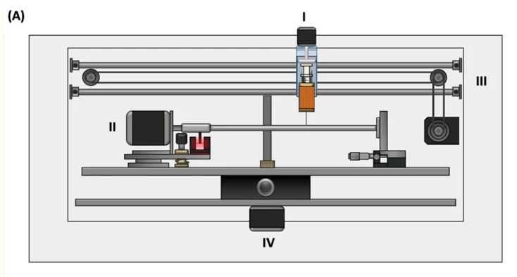
Motor-controlled mandrel, III) Motor-controlled z-axis, and IV) movable stage.
The team built a three-axis rotary 3D bioprinter designed specifically for printing tubular vascular constructs. The diagram on page 3 shows the printer layout and the printing concept: a syringe-based extrusion system deposits cell-laden bioink helically onto a rotating rod whose diameter matches the intended inner diameter of the graft. The same figure also illustrates the workflow from liquid bioink mixing to gelation, thrombin crosslinking, and subsequent tissue culture.
This printing strategy is important because it complements cylindrical tissue fabrication better than a standard flat Cartesian setup. It also supports customized graft dimensions more directly, which is one of the practical strengths of the method.
Why the gelatin-fibrinogen bioink mattered
Fibrinogen is attractive for vascular tissue engineering because it promotes de novo collagen synthesis, but it is not readily printable on its own because of its low viscosity. The central innovation in the paper was to blend fibrinogen with gelatin to create a more printable bioink with shear-thinning behaviour suitable for rotary bioprinting.
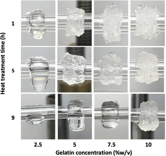
The authors found that heat treatment of gelatin had a major effect on the rheological behaviour of the blend. Longer heat treatment reduced viscosity and storage modulus, while gelatin concentration also strongly affected printability. This made the gelatin fibrinogen bioink tunable, but also made formulation choice critical.
Heat treatment and cell density shaped printability
One of the most useful parts of the study is the formulation detail. The authors showed that printability depended on three main factors: gelatin concentration, duration of heat treatment, and the density of cells mixed into the bioink. The figures in the paper show that extended heat treatment reduced gel structure, while higher cell density also made the bioink more liquid-like and less able to hold shape on the mandrel.
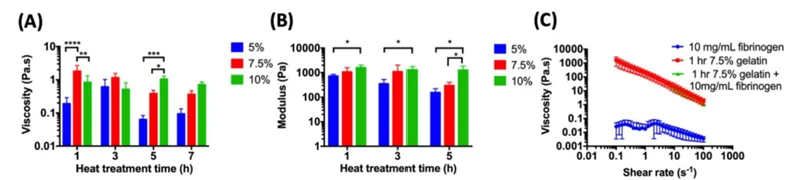
That matters because bioink for vascular constructs is not only about cell compatibility or chemistry. It is also about whether the material can actually hold its structure during printing and immediately afterward. The paper does a good job showing that printability is a balance between rheology, viability, and scaffold formation.
For a related bioink formulation story, see GelMA bioink mechanics for 3D bioprinting.
How the CellScale UStretch was used
CellScale’s role in the paper came through tensile testing of the printed constructs using a CellScale UStretch. The researchers cut strips from the printed vessels in both circumferential and axial directions and tested them in PBS until rupture. Force and displacement data were converted into engineering stress and strain, and these results were then used to calculate elastic modulus, ultimate tensile strength, anisotropy index, compliance, and estimated burst pressure.
This is key because tensile testing of vascular grafts was central to validating the constructs. The study was not just about printing a tube. It was about showing that the mechanical properties improved during culture in ways relevant to vascular function.
For another mechanically focused vascular post, see mechanical properties of heart valve leaflets.
The constructs matured mechanically during culture
The mechanical results are one of the strongest parts of the paper. Over two months of culture, circumferential elastic modulus and ultimate tensile strength increased, while compliance decreased as the constructs matured. The graphs on pages 10 and 11 show this clearly, including the increase in circumferential modulus and burst pressure over time.
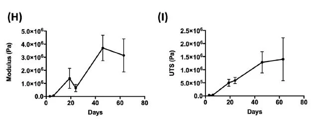
The paper also reports that collagen deposition increased during culture, which likely contributed to the improved mechanics. Histology on page 10 showed stronger matrix accumulation at later time points, while the fluorescence images showed fibroblasts becoming more elongated and organized within the construct.
Burst pressure reached a meaningful benchmark
One of the most valuable translational points in the paper is the burst pressure result. The authors estimated a burst pressure of 1110 mmHg, which they note is about 52% of the value of a human saphenous vein. That does not mean the constructs were fully equivalent to native grafts, but it does show meaningful progress for a printed fibrin-based vascular construct.
This is a strong message for vascular graft mechanical testing. Printing alone is not enough. The real question is whether the construct develops toward mechanically relevant performance, and this study shows that culture-driven maturation pushed the printed vessels in that direction.
Why this study matters for vascular tissue engineering
The broader value of the paper is that it links printing, formulation, and functional mechanics. It shows that a favorable vascular biomaterial such as fibrinogen can become printable by blending it with gelatin, that bioink properties can be tuned through heat treatment and cell density, and that the resulting constructs can undergo ECM-driven mechanical maturation during culture.
For vascular bioprinting, this is exactly the kind of study that matters. It moves beyond proof-of-concept printing and asks whether the printed construct can become a tissue with increasingly relevant mechanical behaviour.
Final thoughts
This paper is a strong example of vascular bioprinting supported by real mechanical validation. The researchers developed a rotary 3D bioprinter and a gelatin-fibrinogen bioink that made fibrinogen printable for vascular construct fabrication. CellScale tensile testing then helped show that the printed grafts gained strength and stiffness during culture, with estimated burst pressure reaching 1110 mmHg.
For readers interested in tissue engineered vascular grafts, bioink design, or mechanical testing of vascular constructs, the study is especially valuable because it connects formulation decisions directly to functional outcomes.
Read the full journal article here: A bioink blend for rotary 3D bioprinting tissue engineered small-diameter vascular constructs
For related reading, you may also like:



