
Hydrogel fibre mechanical testing is rarely as simple as placing a sample in a fixture and pressing start. With hydrated fibres, the surrounding water is part of the experiment. A strand that holds its shape in air can swell, soften, stiffen, or become harder to handle once it is sitting in a bath. That is part of what makes a recent ACS Omega study on recombinant hagfish intermediate filament hydrogel fibres interesting.
In the study, researchers from Utah State University produced protein-based hydrogel fibres from recombinant hagfish intermediate filament proteins, then used the CellScale MicroTester G2 to measure their hydrated mechanical behaviour in a submerged test setup. The paper lands in a useful middle ground: it is about a biomimetic material, but much of the story is really about how to handle and measure a very small wet fibre.
It is not a conventional tissue engineering study. There are no cells seeded onto the fibres and no biological response data yet. Instead, the work asks a more basic mechanical question: when a fibre is thin, hydrated, and sensitive to its bath chemistry, how do you test it without taking it out of the conditions that shape its behaviour?
The answer, in this case, involved a custom submerged bending setup, careful mounting, and mechanical calculations based on fibre deflection.
Why Protein-Based Hydrogel Fibres Need Mechanical Characterization
Protein-based hydrogel fibres can be awkward materials to work with. They are mostly water, they change when the bath changes, and small differences in processing can show up later as differences in stiffness or failure. That is why the mechanical testing matters here. The chemistry gives the fibre a starting point, but it does not tell you how that fibre will behave once it is swollen and mounted in a test fixture.
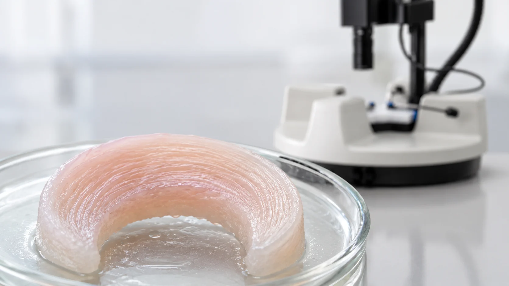
In this paper, the authors are looking at a material that could fit into several different soft-material spaces: tissue engineering, drug delivery, biofabrication, maybe even flexible biomaterial systems. Those applications are still downstream. The immediate question is simpler and more useful for this study: after these recombinant proteins are spun into hydrated fibres, what do the fibres actually do under load?
This is where mechanical characterization of protein hydrogels becomes more than a final checkbox. For hydrated fibres, the test has to deal with small dimensions, delicate handling, and the fact that the material may only behave normally when it remains wet.
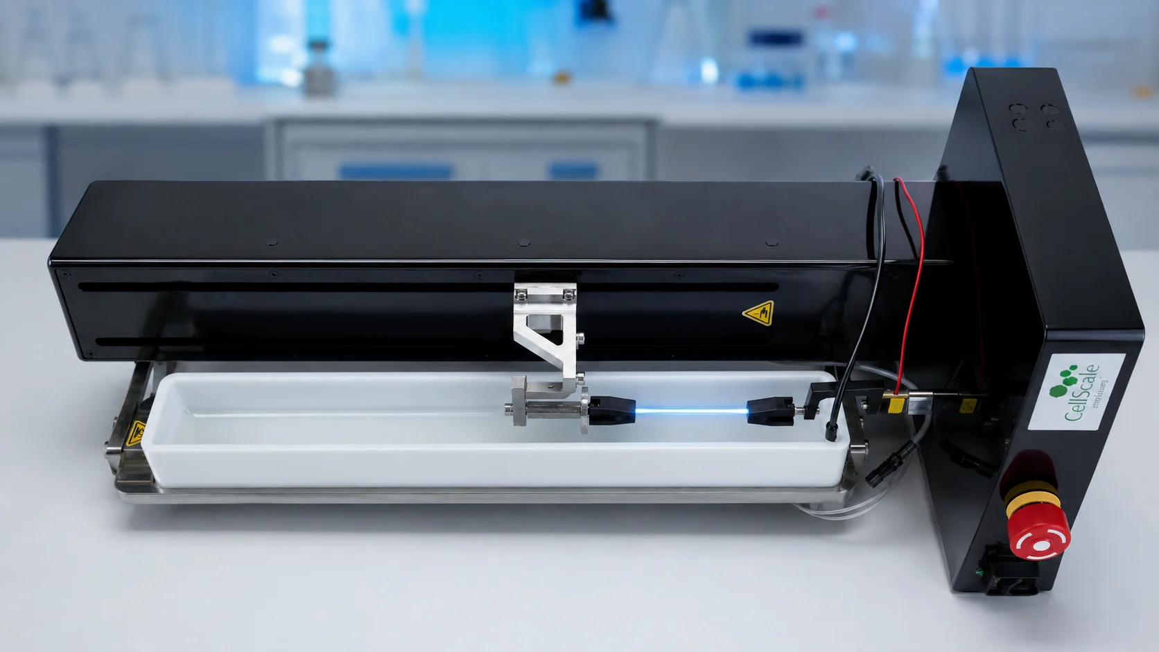
The material in this study came from an unusual biological reference point: hagfish slime threads. In the animal, hagfish intermediate filament threads are deployed into seawater as part of the slime system, so the hydrated environment is not incidental. It is part of the natural context for the material. That makes them a useful reference point for biomimetic hydrogel fibres.
The researchers did not extract fibres directly from hagfish slime for this work. They used recombinant hagfish intermediate filament proteins, which gives the study a different angle. For protein-based biomaterials, making enough material can be just as limiting as the mechanics. A material can have interesting behaviour, but if the protein is difficult to express or purify, it becomes much harder to study at useful quantities.
Spinning Recombinant Protein Hydrogel Fibres from Hagfish Intermediate Filaments
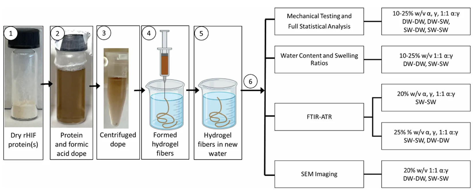
The researchers began with purified recombinant hagfish intermediate filament proteins and dissolved them in formic acid. From there, the protein solution was centrifuged and extruded through a syringe needle into either deionized water or artificial saltwater coagulation baths. After spinning, the fibres were transferred to fresh water baths before testing or characterization.
That processing route produced recombinant protein hydrogel fibres that could be compared across several conditions. The authors varied protein concentration, protein composition, coagulation bath chemistry, and testing bath chemistry. In practical terms, this meant asking whether the same base protein system could produce different hydrogel mechanical properties depending on how it was formed and tested.
One thing that stands out in the workflow is that the fibres were not treated as dry fibres that happened to absorb water later. Hydration was part of the system from the point of coagulation through to mechanical testing. That is an important distinction for hydrogel fibre mechanical testing, because drying or overhandling can shift the material away from the state the researchers are actually trying to measure.
Hydrogel Fibre Mechanical Testing Using a Submerged Bending Setup
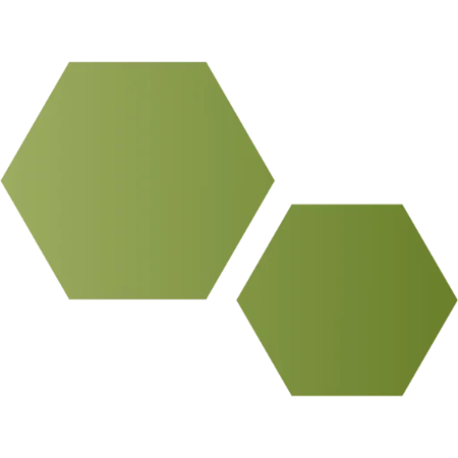
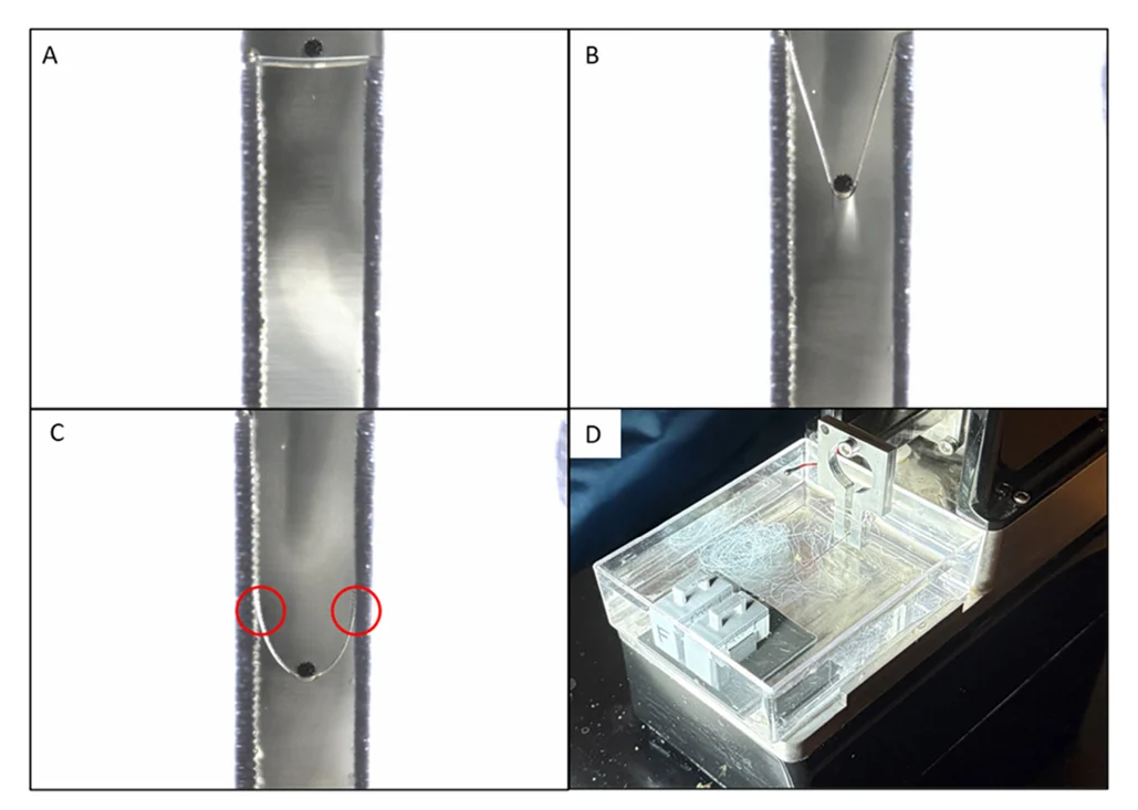
For researchers asking how to test hydrogel fibres mechanically, this paper gives a useful example. The authors used a MicroTester G2 with a bath, a tungsten beam, and a custom 2 mm spacer to perform submerged testing on individual hydrogel fibres.
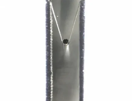
Each fibre was placed across the spacer gap and held with custom clamps. The fibre had to be visibly taut, but not pre-stretched. The MicroTester beam was positioned above the fibre and moved downward at a controlled rate. As the beam displaced the fibre, the system recorded force and displacement. The test continued until the fibre broke or the planned test endpoint was reached.
This is not the same as gripping a dry fibre in a conventional tensile testing frame. The specimen was small, wet, and supported in a geometry where bending and fibre extension occurred together. The authors then used the measured force, displacement, fibre diameter, and gap geometry to calculate stress, strain, elastic modulus, and energy to break.
The material was tested while submerged, with the bath condition matching the second part of each test label. For example, fibres could be formed in saltwater and tested in saltwater, or formed in deionized water and tested in deionized water. That allowed the researchers to look at the role of water chemistry, not just protein formulation.
The images from the MicroTester setup are helpful because they show how delicate three-point tensile tests are. The fibre is thin, the contact region is small, and visual alignment matters. With soft hydrated materials, it is easy for the mechanical result to reflect mounting issues rather than material behaviour. Here, the custom spacer and clamps gave the authors a repeatable way to hold the fibre across a known gap.
Three-Point Bending Test Geometry for Hydrogel Fibres
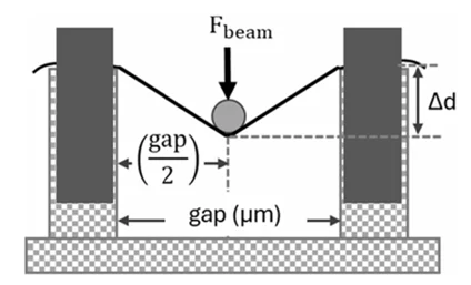
The authors include a diagram of the three-point bending fixture on the MicroTester, explaining how the submerged test geometry worked.
The fibre was secured across two support towers, while the beam pressed downward near the centre. As the beam moved, the fibre formed a triangular shape between the supports and contact point. The authors used the vertical displacement, support gap, force, and fibre diameter in their calculations.
If you’re interested to learn more about bending tests, go to our Flexural & Bending Testing page.
For small, compliant fibres, a setup like this also makes hydrated fibre testing more realistic. The fibre can remain in water, the test can be observed through the imaging system, and the fixture can be scaled around the specimen instead of forcing the specimen into a larger test format.
How Water Chemistry Changed Hydrogel Mechanical Properties
The hydrogel fibre mechanical testing results showed that the fibres were highly tunable. Depending on protein composition, protein concentration, and bath condition, the reported elastic modulus values ranged from about 106 to 1041 kPa. Stress, strain, and energy to break also changed across groups.
The saltwater conditions were especially interesting. In several cases, fibres formed and tested in saltwater showed stronger or more consistent mechanical behaviour than comparable deionized water conditions. The authors connect this partly to ionic crosslinking from divalent ions in artificial seawater, such as magnesium and calcium. That interpretation makes sense, although the exact relationship between water chemistry, swelling, structure, and mechanics is not something to reduce to one variable.
For the alpha fibres, increasing protein concentration from 20% to 25% in saltwater increased stress, strain, energy to break, and elastic modulus. The 1:1 alpha/gamma fibres also showed a concentration-dependent increase in stress and modulus under saltwater conditions. The gamma fibres behaved a little differently, with less improvement at higher concentration. That unevenness is useful. It suggests that the protein composition was not just a label on the sample. It affected how the fibre responded.
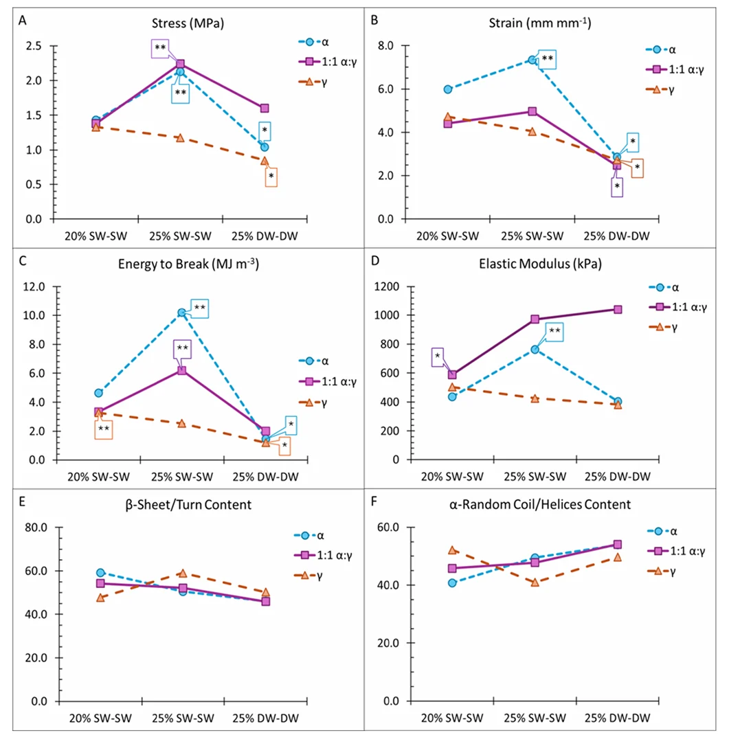
The mechanical testing of hydrated hydrogel fibres also showed that deionized water conditions could reduce strain and energy to break, even when modulus was still relatively high in some groups. In practice, that means stiffness alone does not tell the whole story. A fibre can be stiff but less extensible, or it can absorb more energy before breaking depending on how it was processed.
Related reading: Hydrogel stiffness measurement across five test methods.
Mechanical Characterization of Protein Hydrogels Is Not Just About Modulus
A tidy summary would be to say that the authors made tunable hydrogel fibres. That is true, but it skips some of the more useful parts of the paper.
The study measured several mechanical outputs at once: stress, strain, elastic modulus, and energy to break. Those values do not always move together. A condition that increases modulus may not give the highest strain. A fibre that reaches higher stress may not have the same energy-to-break behaviour as another formulation. For protein-based hydrogel mechanical properties, that matters because the best material depends on what the fibre is supposed to do.
A hydrogel fibre intended for a soft scaffold, a biofabrication process, or a flexible material system may not need the same stiffness or failure behaviour. This is one reason microscale mechanical testing of soft fibres is useful. It lets researchers work with the actual sample format, rather than casting a larger bulk gel and assuming the fibre version behaves the same way.
What the MicroTester Was Doing in This Study
In this publication, the MicroTester was used as a small-sample mechanical testing platform for individual hydrated fibres. The researchers used the instrument bath to keep samples submerged, the three-point tensile testing fixture for flexural testing, the microbeam to apply controlled displacement, and the software to collect force and displacement data during testing.
The setup also relied on imaging. The authors measured hydrated fibre diameter using the MicroTester software, and the test progression images show the fibre before loading, during deflection, and at break. For small hydrated samples, that visual record is not just a nice add-on. It helps confirm contact, alignment, mounting, and failure behaviour.
The authors then exported the data and calculated stress, strain, energy to break, and elastic modulus using the measured diameter and the geometry of the support gap. That workflow is a good example of how micro-scale mechanical testing often works in research: the instrument provides controlled motion, force measurement, imaging, and environmental support, while the analysis is adapted to the sample geometry.
Why Hydrated Mechanical Testing Matters for New Biomaterials
For new biomaterials, especially hydrogels and soft protein systems, the environment can be part of the material. Water content, ion concentration, swelling state, and test geometry may all affect what gets measured.
That is the larger lesson from this study. Hydrogel fibre mechanical testing was not just used to assign a modulus value to a new material. It helped the researchers compare how recombinant protein composition, concentration, and water chemistry shaped the fibre response. The same general material system could produce different behaviour depending on how it was spun and where it was tested.
There is still work ahead before recombinant hagfish intermediate filament hydrogel fibres can be treated as application-ready biomedical materials. The authors point toward future biocompatibility studies and more detailed structural work. That seems reasonable. At this stage, the paper is strongest as a mechanical and structural characterization study of a new protein-based hydrogel fibre platform.
Still, the work is a useful example for anyone developing hydrated fibres, soft biomaterials, or small filament-like samples. When the material is small, wet, and sensitive to its surroundings, the mechanical test has to be built around that reality.
Citation
Tailorable Hydrogel Fibers from High-Yield Recombinant Hagfish Intermediate Filament Proteins: A New Frontier in Biomimetic Materials
Brianne E. Bell, Oran Wasserman, Thomas I. Harris, Hayden B. Johns, Paula E. Oliveira, and Justin A. Jones
ACS Omega 2026 11 (23), 33619-33629
About the MicroTester
The CellScale MicroTester is used for low-force mechanical testing of small and delicate samples, including hydrogels, microtissues, fibres, thin materials, and other soft biomaterials. For hydrogel fibre mechanical testing, the main practical advantage is that the sample can be tested at the scale where it actually exists.
In studies like this one, that means researchers can work with individual hydrated fibres rather than reshaping the material into a larger specimen. The test can be performed in a bath, observed through integrated imaging, and adapted with custom fixtures when the specimen geometry calls for it.
For researchers working on hydrogel mechanical testing, biomimetic hydrogel fibres, or other small soft material systems, the study is a useful example of how mechanical testing can be adapted without removing the material from its hydrated state.