


Investigating regional variations in the biaxial mechanical properties of heart valve leaflets

A tensile test divider icon

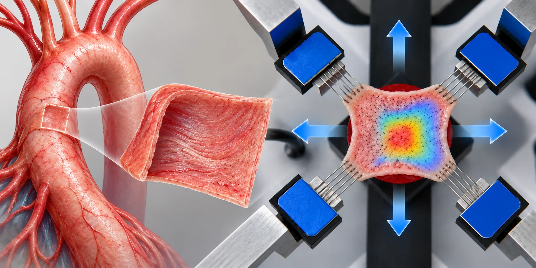
Dr Chung-Hao Lee and his cardiovascular biomechanics team in the School of Aerospace and Mechanical Engineering at the University of Oklahoma uses the CellScale BioTester to further research on porcine atrioventricular heart valve leaflets.

CellScale hexagon shapes

Heart valve disease is a debilitating disease that affects millions of people. It occurs when one or more of the four valves in the heart does not open or close properly. Mechanical characterization of heart valve leaflets is critical for improving therapeutic options for valvular disease. Many attempts at characterization focus on the mechanical properties of the central region and neglect spatial variation in the tissue’s properties.

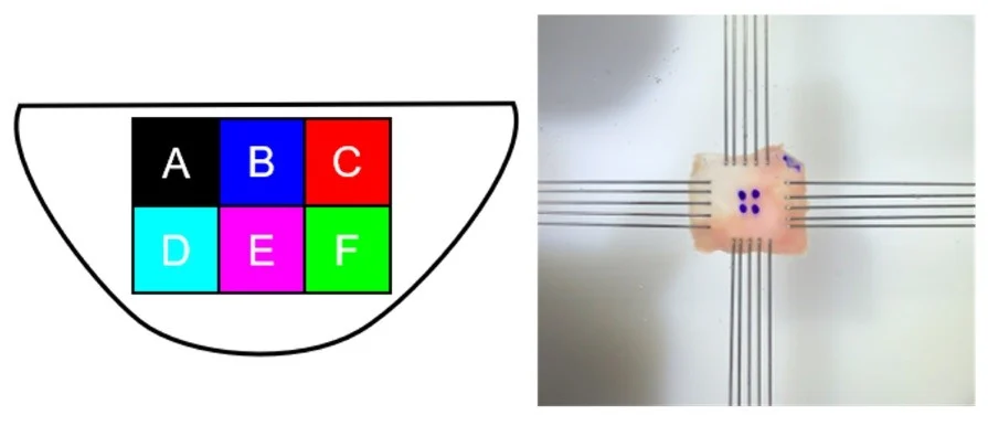
Dr Lee and his team at the University of Oklahoma have utilized the CellScale BioTester to mechanically characterize six distinct regions in heart valve leaflet tissues. Specifically, they have determined that anisotropy and peak stretch vary between regions and that these variations are important for proper leaflet health and function.

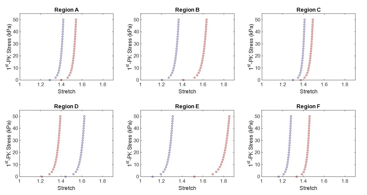
Experimenting on both porcine mitral valve and tricuspid valve anterior leaflets, it was discovered that (i) central regions of the leaflet were more anisotropic than edge regions, (ii) the mitral valve anterior leaflet extended more closer to the perimeter, and (iii) mitral valve leaflet tissue regions exhibited a greater decay than tricuspid valve regions in stress-relaxation behavior. Results plotted of the Stress against Stretch for each of the 6 regions are shown below.
Read the full journal article here: https://doi.org/10.1016/j.jbiomech.2018.11.015

Read about another user publication highlight (3D bioprinting of human tissue with dECM) here.

CellScale hexagons, without text

CELLSCALE INSTRUMENT USED

TAGS

Biaxial mechanical testing, BioTester, Cardiovascular Biomechanics, Heart Valve Mechanics, Mitral Valve, Stress Relaxation, Tissue Anisotropy, Tricuspid Valve

POST DETAILS
CATEGORY

Research Highlights

INSTRUMENT USED
BioTester
RESEARCH APPLICATIONS
Cardiac Tissue Engineering & MechanicsHeart Valve Tissue Engineering & Mechanics
TESTING METHODS
Biaxial TestingStress Relaxation TestingViscoelastic & Time-Dependent Testing

Related Posts:

Filter by Category:
Filter by Instrument
Filter by Testing Method:
Filter by Research Application:
Bioprinted meniscus tissue engineering construct with curved collagen-like alignment patterns in the foreground and a softly blurred CellScale BioTester in the background.

July 2, 2026

Meniscus Tissue Engineering with Boundary-Guided Collagen Alignment

Hydrogel fiber mechanical testing setup showing a thin hydrated fibre suspended across two supports while a small MicroTester beam presses downward in the centre, before and after.

June 25, 2026

Hydrogel Fibre Mechanical Testing of Recombinant Hagfish Protein Materials

MSC spheroids embedded in a translucent PEG-fibrin hydrogel within soft vocal fold-like tissue, with a blurred CellScale MicroTester in the background.

June 11, 2026

Vocal Fold Regeneration with MSC Spheroids

Hydrogel eye drop on a corneal surface with a mechanical testing probe and force-distance curve representing mucoadhesion testing for ocular drug delivery.

May 28, 2026

Mucoadhesion Testing of Hydrogel Eye Drops for Ocular Drug Delivery

May 14, 2026

Flexible Flow Sensor Testing for Pioneering Autonomous Robotics

Featured illustration showing a layered tissue phantom under compression, an elastography heatmap, a phase velocity versus depth graph, and an AI neural network used to support depth-resolved stiffness analysis.

May 7, 2026

Validating AI-Based Elastography Measurements with Compression Testing

Contact Sales

Product of Interest:
CellScale hexagon shapes