
Corneal Biomechanics
and Ophthalmic Tissue Engineering

Overview of Corneal Biomechanics and Ophthalmic Tissue Engineering
Ophthalmic biomechanics focuses on the mechanical behaviour of ocular tissues that must preserve shape, transparency, and load-bearing function under physiologic pressure and external forces. The cornea is a thin, collagen-rich, layered structure whose mechanical response is strongly influenced by stromal lamellae orientation, hydration state, and crosslink density.
- Mechanical characterization supports research in:
Corneal tissue engineering aims to develop biomimetic stromal equivalents, regenerative scaffolds, and hydrogel-based corneal substitutes that reproduce native mechanical and optical behaviour. Publications frequently compare engineered constructs against native corneal tissue using uniaxial, biaxial, and low-force testing methods under hydrated conditions.

Importance of Mechanical Testing in Corneal Biomechanics Research
Across published corneal biomechanics studies, mechanical testing is commonly used to resolve anisotropy, quantify stiffness changes associated with disease or crosslinking treatments, and evaluate regional mechanical heterogeneity across the corneal dome. Subtle alterations in corneal stiffness and viscoelasticity have been linked to refractive stability, ectasia risk, and surgical response.
Mechanical testing enables researchers to:
- Quantify corneal stiffness changes associated with disease, remodeling, or hydration shifts
- Measure directional mechanics linked to stromal collagen alignment
- Evaluate mechanical stability following crosslinking or scaffold fabrication
- Map localized stiffness gradients that influence curvature and optical performance
- Benchmark engineered corneal constructs against native tissue mechanics
- Assess time-dependent behaviour relevant to intraocular pressure loading
- Generate parameters for computational models of ocular mechanics and surgical planning
Accurate corneal mechanical testing improves reproducibility and strengthens structure-function interpretation in both basic and translational ophthalmic research.
Recommended CellScale Instruments for Corneal Mechanical Testing
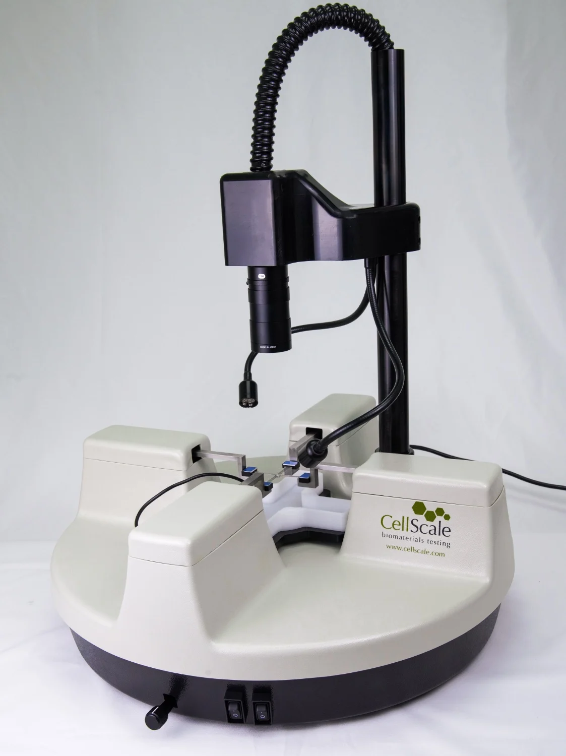
BioTester
Used for biaxial testing of thin, planar ocular tissues and engineered corneal constructs where anisotropy and in-plane strain behaviour are central to corneal biomechanics.
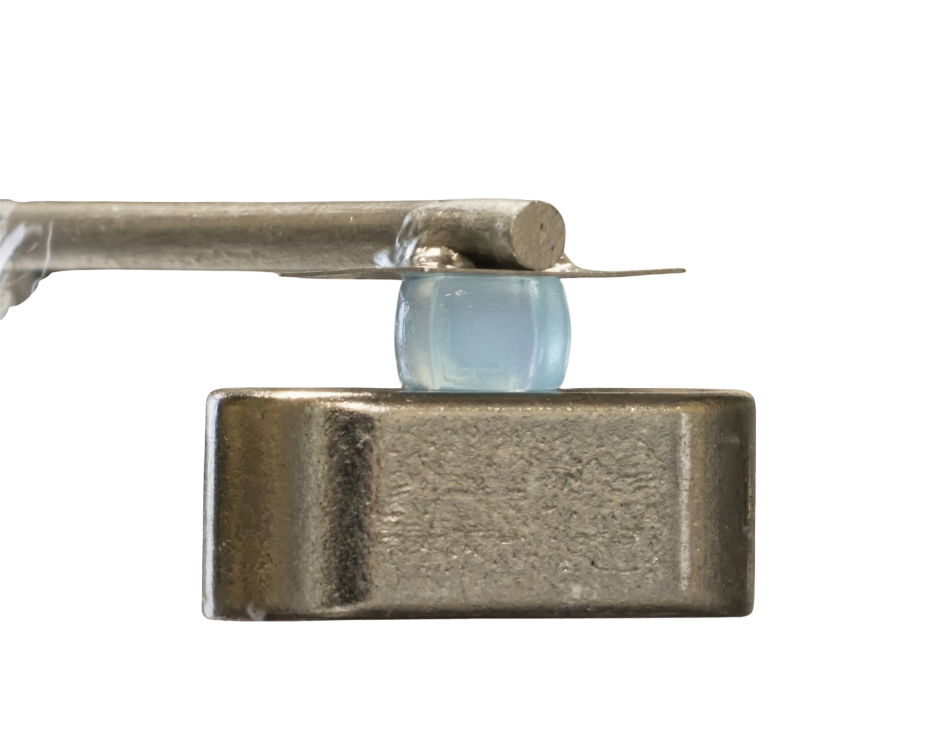
MicroTester
Ideal for low-force indentation, micro-compression, and localized stiffness mapping of corneal tissues and small engineered constructs under hydrated conditions.
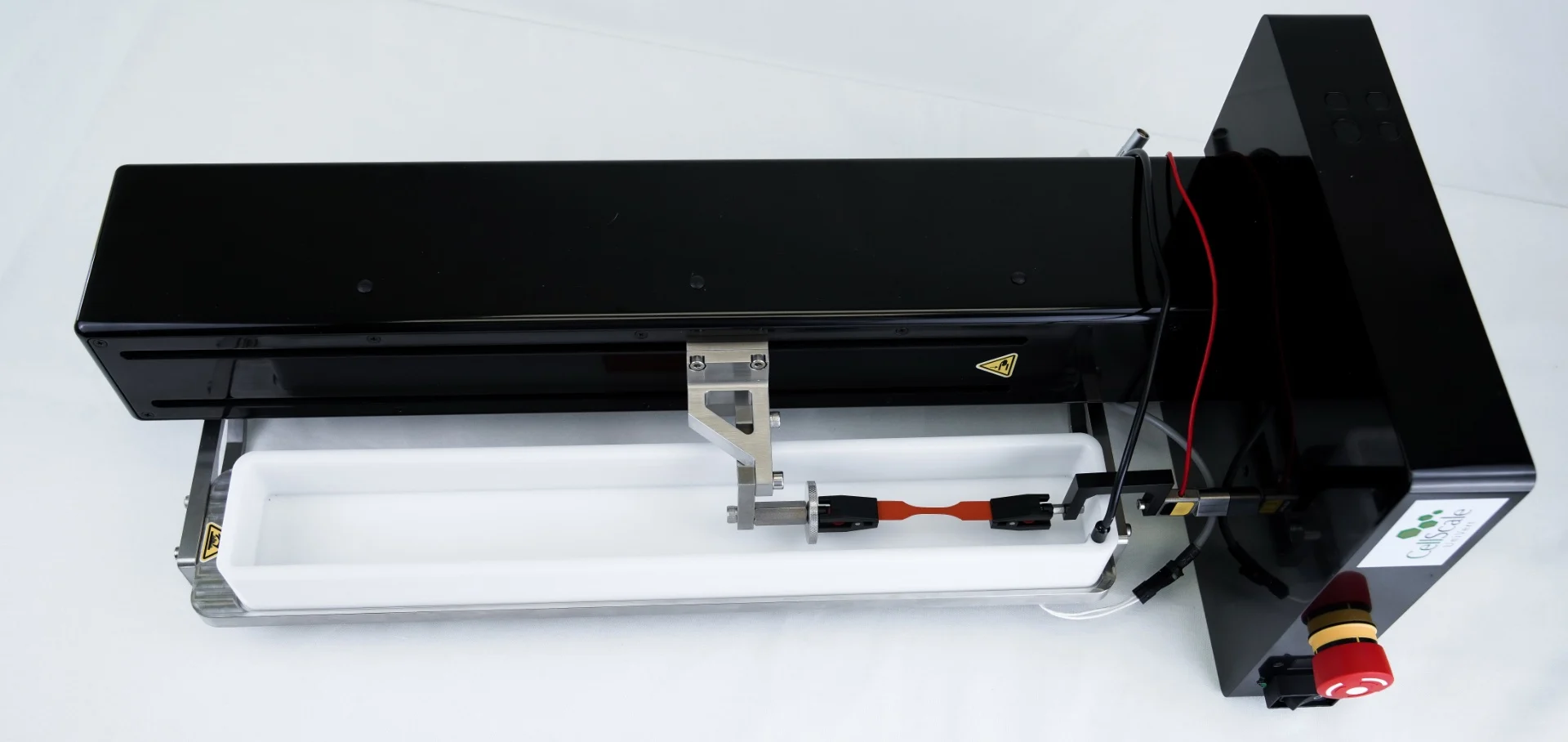
UniVert
Supports uniaxial tensile testing of corneal strips, scleral samples, and corneal scaffolds across a wide range of stiffnesses using sensitive force resolution.
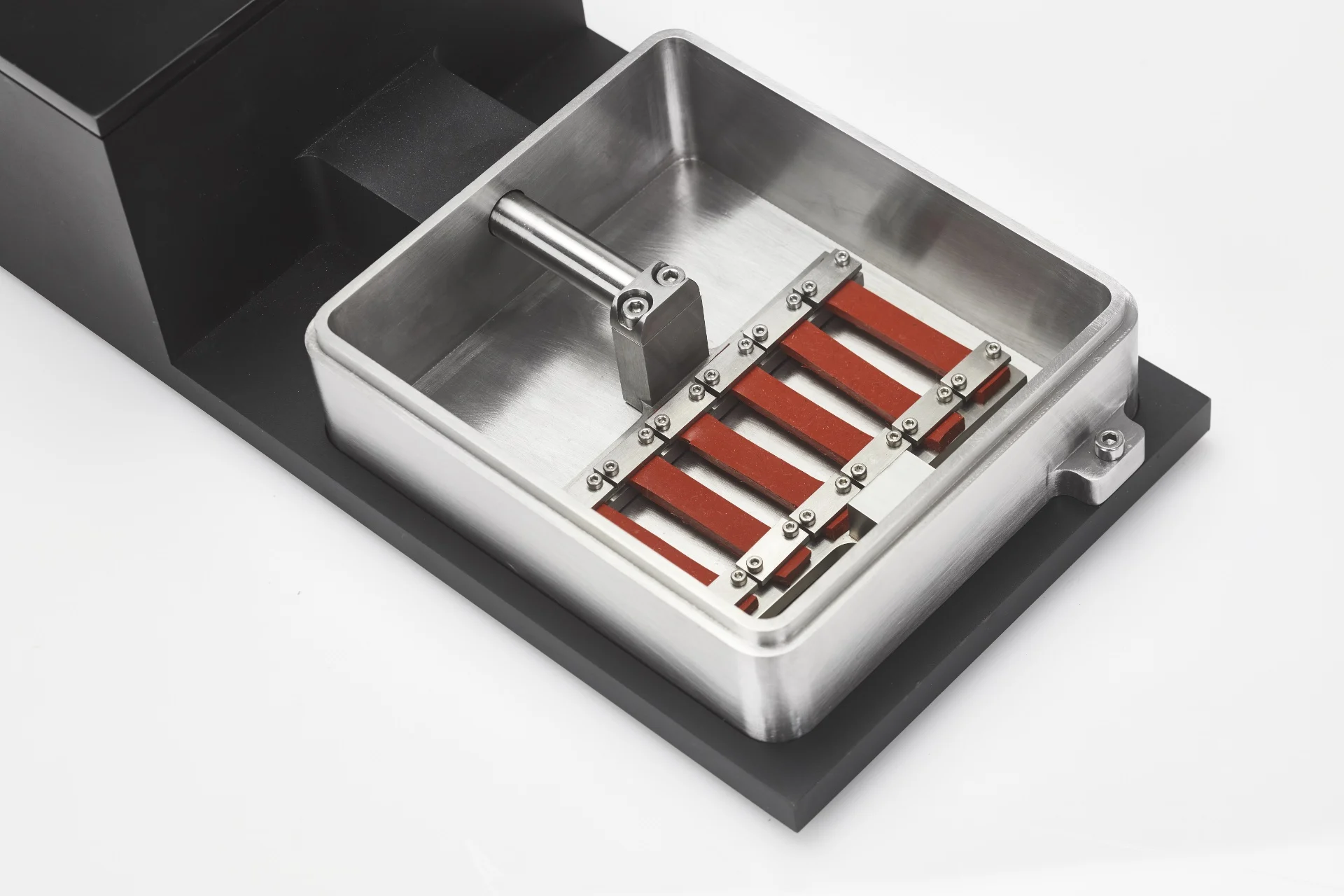
MechanoCulture T6
Applies controlled cyclic stretch to engineered ocular tissues or biomimetic membranes to investigate mechanobiology, remodeling, and strain-dependent matrix adaptation.
Testing Methods for Corneal Biomechanics and Ophthalmic Tissue Engineering
Indentation Testing
Maps corneal stiffness and regional heterogeneity across the ocular surface
Biaxial Testing
Evaluates corneal anisotropy and multiaxial in-plane mechanical response
Stress Relaxation Testing
Measures time-dependent stress dissipation in soft, hydrated biological materials
Digital Image Correlation
Measures full-field strain and deformation patterns during corneal loading
Flexural & Bending Testing
Measures bending stiffness of corneal and ocular tissues relevant to shape stability
Representative Sample Types in Ophthalmic Biomechanics
Native ocular tissues
- Cornea and stromal layers
- Sclera
- Conjunctival tissues
Engineered corneal tissue engineering constructs
- Collagen-based stromal equivalents
- Decellularized corneal matrix scaffolds
- Hydrogel corneal substitutes
- Aligned fibre-reinforced corneal constructs
Ophthalmic biomaterials and device-related samples
- Contact lens and ophthalmic hydrogel materials
- Corneal adhesives and sealants
- Thin films and membrane-based ocular interfaces
Disease and intervention models
- Crosslinking-modified corneal tissues
- Ectasia and keratoconus-inspired mechanical models
- Remodeling and fibrosis-associated ocular tissues
Selected Publications in Corneal Biomechanics
Advance Your Corneal Biomechanics and Ophthalmic Tissue Engineering Research
CellScale systems support corneal biomechanics measurement, corneal mechanical testing, and ophthalmic tissue engineering studies requiring sensitive force control and physiologic hydration conditions. Contact our team to identify the ideal platform for your ocular tissue mechanics workflow.