A bioink needs to do more than produce a defined 3D printed structure… it also needs to support the living cells inside it.
But what happens when the printing conditions that produce the most accurate structures aren’t the ones that give cells the best environment?
Researchers at Friedrich-Alexander-Universität Erlangen-Nürnberg (FAU) in Germany screened alginate dialdehyde-gelatin (ADA-GEL) formulations for C2C12 mouse myoblasts. Their investigation of bioink mechanical properties included compression measurements using a CellScale MicroTester LT over 14 days, rheology, and a series of printing and cell experiments.
The researchers found that better print accuracy did not necessarily correspond to more favourable cell responses. Their measurements also showed that the hydrogels became substantially softer after immersion, raising questions about when bioink mechanical properties should be measured during material development. This is relevant to researchers working on 3D bioprinting and bioink materials testing.
Six ADA-GEL Bioink Formulations Narrowed Down
The low, medium and high compositions contained 2.5% ADA + 3.75% gelatin, 2.5% ADA + 5% gelatin, and 3.75% ADA + 7.5% gelatin, respectively (all weight/volume). Each was prepared with gelatin dissolved either at 37°C for 15 minutes or at 80°C for three hours.
This produced six inks from three polymer compositions, with gelatin preparation temperature as the second variable.
The 80°C low ink couldn’t maintain a stable multilayer grid. Both high compositions retained shape better but needed pressures exceeding 140 kPa. That is a printing requirement to weigh carefully before adding cells.
Inks made with gelatin prepared at 37°C had higher storage and loss moduli and complex viscosity than matching 80°C preparations. The authors discuss changes in gelatin chain characteristics following the longer, hotter treatment. Swelling, degradation and C2C12 surface cultures supplied further selection criteria.
The medium pair emerged as the most favourable combination of printability, handling and cell compatibility, and was renamed 37_ADAGEL and 80_ADAGEL.
How Did the MicroTester LT Measure Bioink Mechanical Properties?
Once the two medium ADA-GEL formulations had been selected, the researchers examined their compressive stiffness. They also prepared versions containing 0.1% (w/v) silicate-based bioactive glass nanoparticles, bringing the comparison to four formulations.
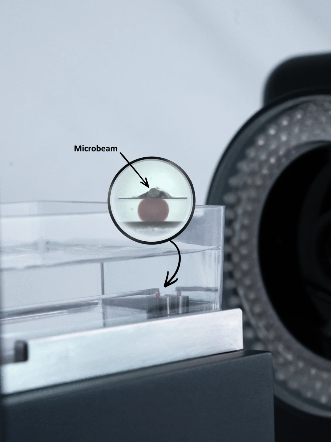
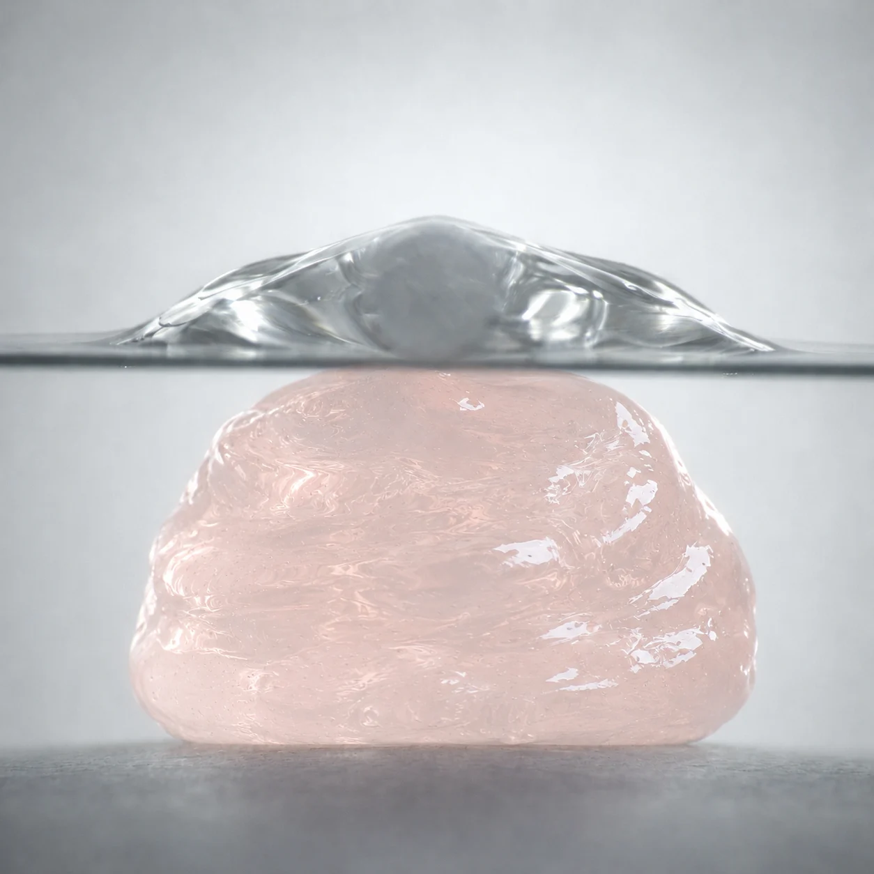
The specimens were small cylinders, 5 mm across and 2 mm high. Each was compressed between 6 × 6 mm platens on a MicroTester, with six specimens tested per condition. The instrument used 60 mm cantilevers, selecting microbeam diameters of 0.3048, 0.4064 or 0.5588 mm to accommodate the forces involved. Effective compressive modulus was determined from the first 5% strain.
The team tested the hydrogels immediately after preparation, then again following 1, 3, 7 and 14 days of incubation. Compression took place in HBSS at 37°C, so the specimens were measured in a warm, hydrated environment rather than dry at room temperature.
Rheology provided a separate set of measurements. For the inks, the researchers ran frequency sweeps at 25°C, using 5% oscillatory strain. Those tests measured shear behaviour, whereas the MicroTester measurements described the effective compressive modulus of the formed hydrogels.
For additional background on these measurements, see CellScale’s hydrogel mechanical testing and hydrogel stiffness measurement resources. Our Research Highlight on GelMA bioink mechanics covers a separate study of formulation-dependent mechanical behaviour.
How Does Hydrogel Stiffness Change During Incubation?
The biggest change in stiffness happened during the first day. Before immersion, the 37_ADAGEL hydrogel had an effective compressive modulus of 3.2 ± 1.0 kPa, and the other formulation prepared with gelatin treated at 37°C also showed higher average stiffness than its 80°C counterpart.
By day 1, all four formulations had become substantially softer. Their effective moduli then changed relatively little through day 14, with no substantial effect from adding 0.1% (w/v) bioactive glass nanoparticles.
The researchers linked the initial decrease to swelling as the hydrogels absorbed fluid. They also discussed the possibility that testing in HBSS at 37°C, rather than at room temperature without immersion, contributed to the lower stiffness. The experiment did not isolate these effects, so the reduction cannot be assigned to swelling or temperature alone.
This difference between the freshly prepared and incubated samples is particularly relevant when comparing hydrogel formulations. A modulus measured on day 0 does not necessarily describe the material after several days in culture conditions. Oxidation degree, crosslinking, strain range and the measurement method also need to be considered when comparing results with other ADA-GEL studies.
Conical and Cylindrical Nozzles at the Same Outlet Diameter
The conical and cylindrical nozzles had the same 250 µm outlet diameter, but the inks behaved differently as they passed through them. The cylindrical nozzle had a narrow channel extending along its length, while the conical nozzle tapered towards the opening.
The difference showed up in both the printed grids and the pressure needed to produce them. Cylindrical nozzles generally gave higher diagonal crossing ratios (DCR), indicating better print accuracy. With cell-free 37_ADAGEL, however, switching from a conical to a cylindrical nozzle increased the required pressure from approximately 70 to 80 kPa to 225 kPa.
The pressure measurements for cell-laden printing were different. For example, the researchers reported 140 kPa for 37_ADAGEL containing myoblasts when printed through a cylindrical nozzle. Those settings should be considered separately from the cell-free grid experiments, rather than treated as measurements under identical conditions.
C2C12 Myoblast Metabolic Activity and Orientation
C2C12 myoblasts were encapsulated at 2 million cells/mL. In every investigated group, WST-8 absorbance rose over time. The highest day-3 and day-7 readings came from the 37_ADAGEL conical group. By day 14, each conical group had significantly higher absorbance than the corresponding cylindrical group.
WST-8 measures metabolic activity and was used here as a proxy related to viable cells. Its absorbance readings should not be taken as direct cell counts or survival percentages.
The day-7 confocal images showed elongated cells in the printed filaments. Phalloidin-labelled actin was analysed relative to the printing direction.
The orientation table above reports mean angular deviations; smaller values indicate closer orientation to that direction.
The 37_ADAGEL conical condition had the lowest mean, but the orientation differences were not statistically significant. The authors also identify cell density and computation method as possible influences on the angles. That supports describing a trend, without claiming an alignment improvement.
A later experiment used conical nozzles to print the nanoparticle-composite bioinks. At day 7, the 37°C-prepared composite had higher WST-8 absorbance and a lower mean angular deviation than its 80°C counterpart.
Selecting a Formulation for Myoblast Bioprinting
The combined assessment favoured the medium formulation made with 37°C-prepared gelatin, printed using a conical nozzle.
Grid fidelity generally favoured cylindrical nozzles, but the cell metabolic activity measurements favoured conical nozzles. At the same time, the compression results showed that the hydrogels became substantially softer after immersion, with relatively stable effective moduli over the remaining incubation period.
The bioink mechanical properties, printing conditions and biological measurements therefore need to be considered together. A stiffness-only explanation of the biological findings would go beyond the experiment, where several material and printing variables differed.
Mature, functional or contractile muscle tissue was not demonstrated. The findings concern formulation selection, myoblast behaviour and hydrogel mechanical characterization during incubation.
For a related example of mechanical characterization in muscle scaffold development, see piezoelectric hydrogels for skeletal muscle tissue engineering.
Original Publication
Hsuan-Heng Lu, Clara Steinert, David Böhringer, Margitta Büchner, Dirk W. Schubert, Sahar Salehi, Aldo R. Boccaccini, Screening ADA-GEL based hydrogel formulations and bioprinting conditions for myoblast encapsulation and alignment, Bioprinting, Volume 60, 2026, e00505, ISSN 2405-8866, https://doi.org/10.1016/j.bprint.2026.e00505.
About the CellScale MicroTester
The MicroTester is designed for mechanical characterization of small, soft and delicate specimens like hydrogels, microtissues, spheroids and engineered biomaterials.
The instrument uses calibrated microbeams to measure force while controlling specimen displacement. Integrated imaging allows researchers to observe and position samples during testing. Depending on the configuration, testing can also be performed with hydrated specimens under controlled temperatures.
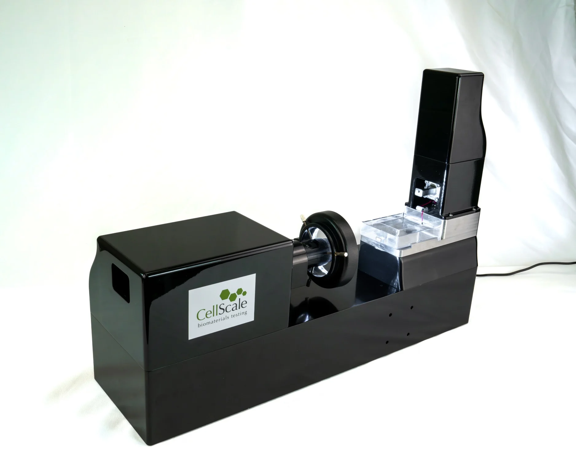
In this study, the researchers used the MicroTester LT to compare the effective compressive modulus of ADA-GEL formulations over 14 days. The MicroTester supports compression, indentation, tension, and shear testing, with sensitive force measurement, integrated imaging, and hydrated and temperature-controlled testing capabilities.
For researchers measuring hydrogel stiffness, the MicroTester mechanical testing system provides sensitive compression and indentation measurements for small, soft specimens, including hydrated materials.