Vascular Graft Mechanical Testing with a Custom Tubular System


University of Pittsburgh
Vande Geest Laboratory | Department of Bioengineering
Project Background
Tissue-engineered vascular grafts are being developed as alternatives to conventional vascular graft materials, particularly for small-diameter blood vessel replacement. Alongside biocompatibility and long-term remodeling, the mechanical behaviour of these constructs is an important consideration. Differences in compliance between an engineered graft and the surrounding native artery may affect local loading conditions and influence how the graft remodels following implantation.
Measuring these properties requires more than conventional uniaxial tensile testing. Blood vessels experience internal pressure while remaining under axial tension, and their diameter changes throughout each pressure cycle. For small specimens, accurately measuring those changes also requires suitable mounting fixtures and optical access.
In 2022, Professor Jonathan Vande Geest’s laboratory at the University of Pittsburgh worked with CellScale to develop a custom mechanical testing system capable of applying controlled axial tension and internal pressure to tubular specimens.
The project followed an earlier collaboration involving a customized MechanoCulture system. This new instrument was designed specifically for mechanical characterization of blood vessels and engineered tubular constructs.
Since its development, the custom testing system has been used in multiple peer-reviewed studies investigating vascular graft material behaviour, compliance matching, hemocompatibility, and tissue remodeling.

Prof. Jonathan P. Vande Geest
Department of Bioengineering, Department of Mechanical Engineering and Material Science, the Department of Ophthalmology, the McGowan Institute for Regenerative Medicine, the Louis J. Fox Center for Vision Restoration, and the Vascular Medicine Institute at the University of Pittsburgh.

The Challenge
Testing Small-Diameter Tubular Specimens
The system needed to accommodate blood vessels and engineered grafts with inner diameters ranging from 0.5 to 6 mm and specimen lengths of approximately 5 to 40 mm.
At this scale, mounting becomes particularly challenging. The specimen must remain secured to its fixtures during inflation without introducing leakage, excessive deformation, or unwanted axial loading.
Applying Axial Tension and Internal Pressure
Vascular tissues respond to combinations of circumferential and axial loading. The laboratory needed to control specimen length or axial force while independently varying internal pressure.
This required a testing configuration with separate loading mechanisms, force and pressure measurement, and enough flexibility to accommodate different experimental protocols.
Measuring Diameter Changes During Inflation
Pressure alone does not describe the mechanical response of a vessel. Its changing diameter must also be measured to calculate circumferential deformation and compliance.
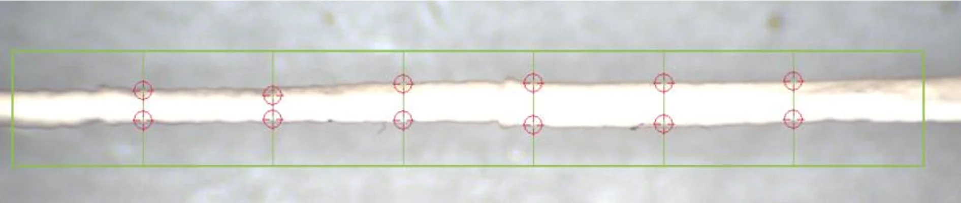
The system therefore required non-contact imaging capable of tracking specimen edges at multiple positions along the vessel as internal pressure increased and decreased.
Maintaining Hydrated Testing Conditions
The researchers also needed to perform mechanical tests with specimens submerged in a temperature-controlled fluid bath, allowing measurements under conditions appropriate for vascular tissue characterization.
Custom Solution Developed by CellScale
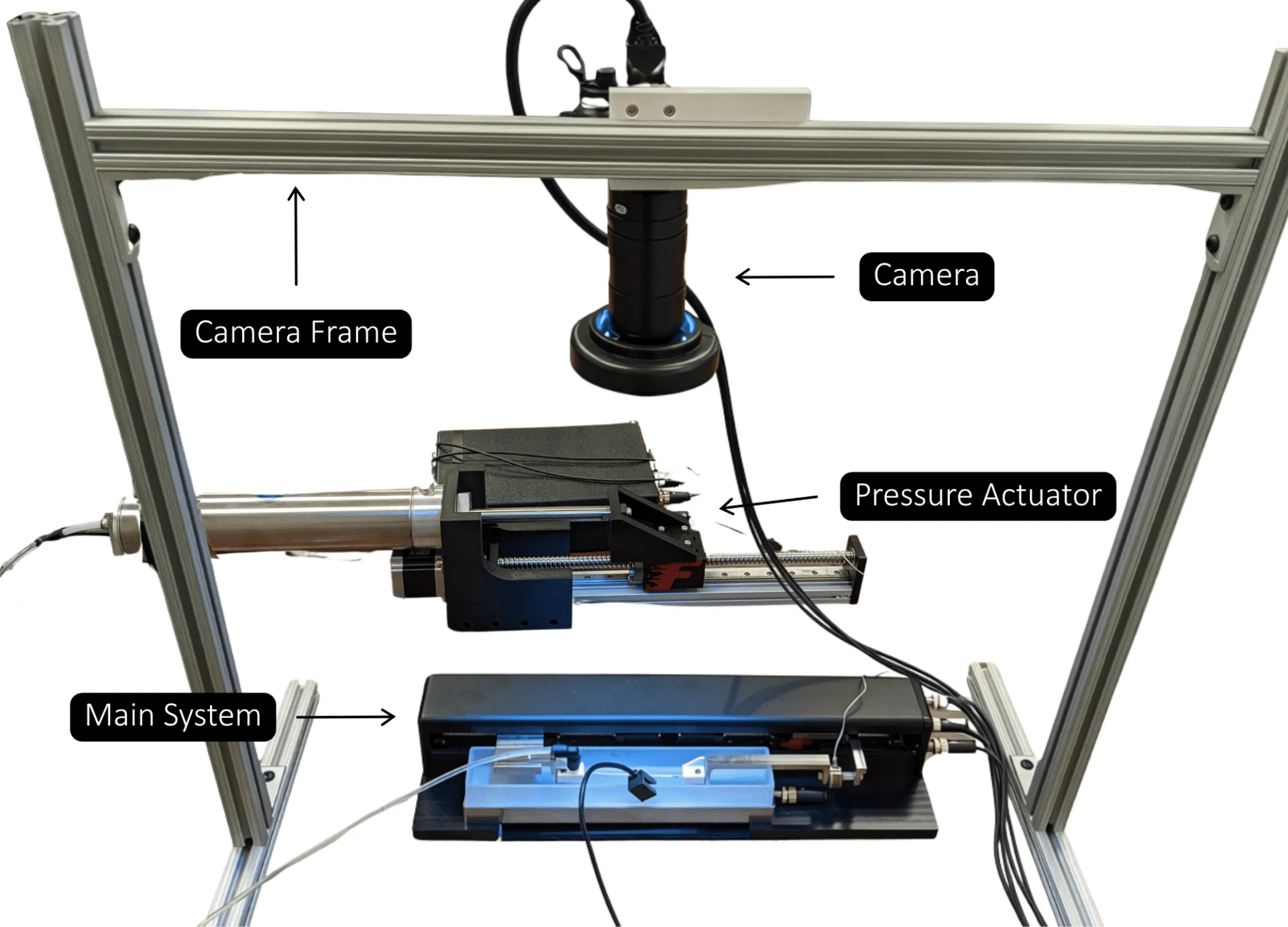
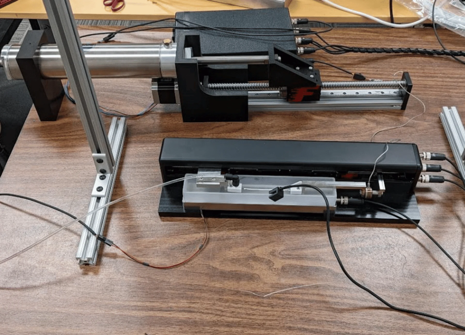
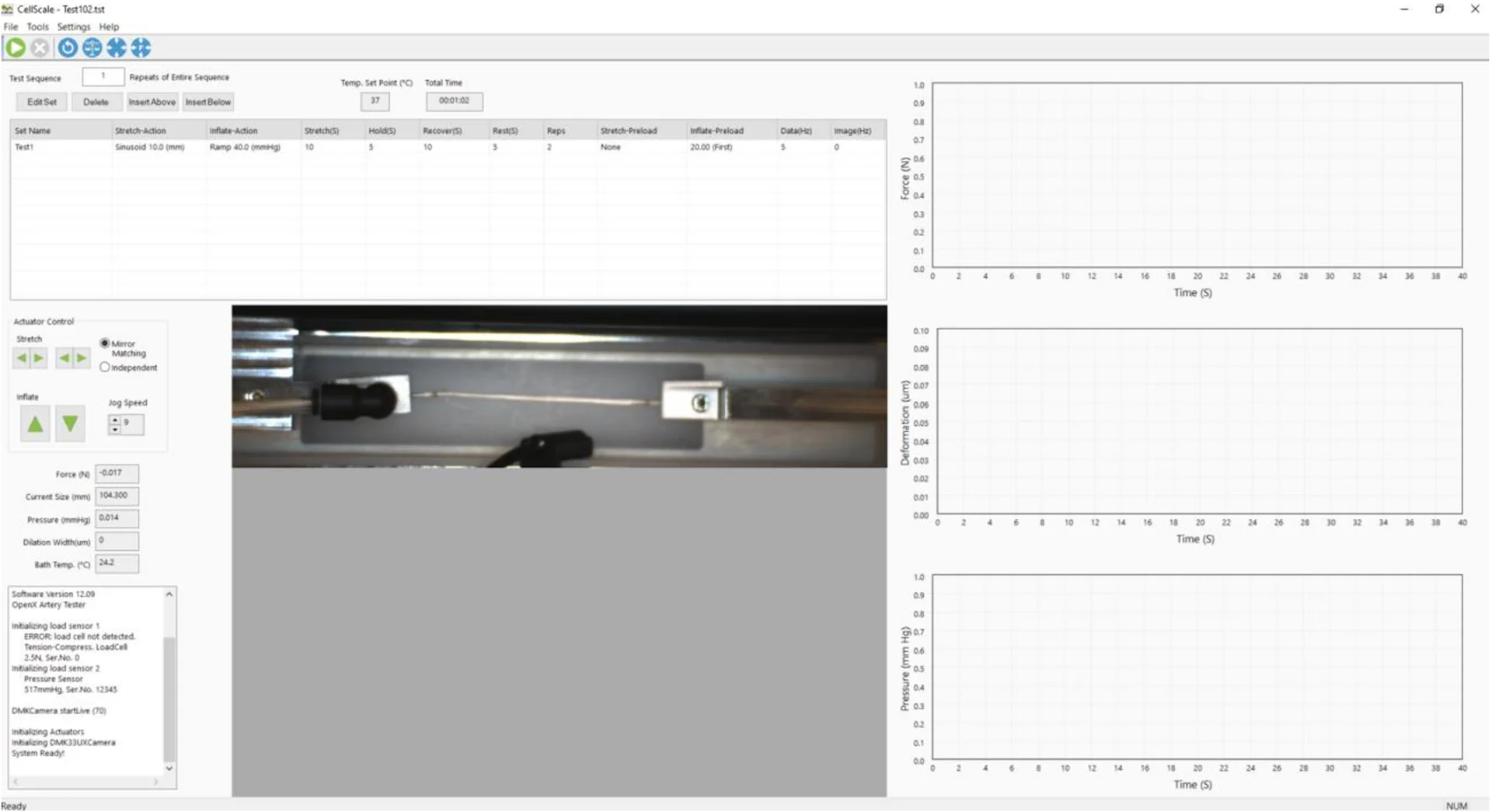
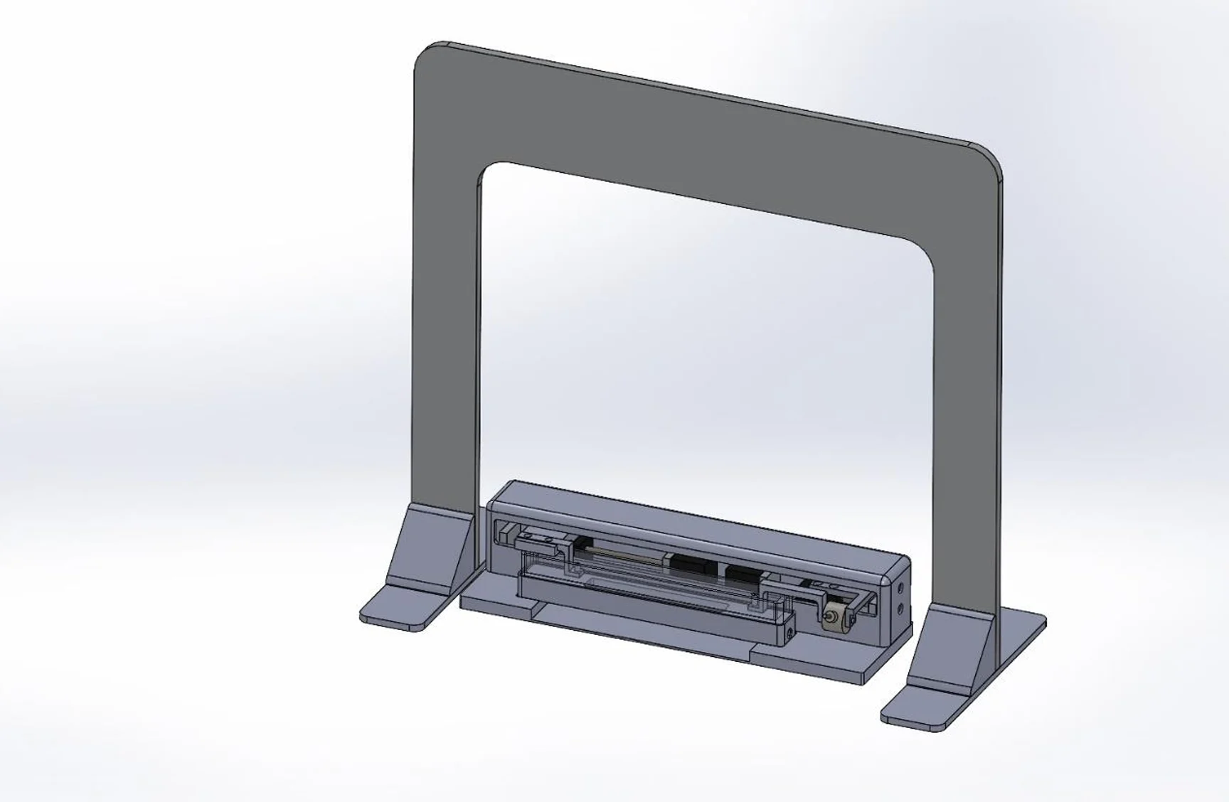
CellScale designed and manufactured a custom tubular mechanical testing system around the laboratory’s experimental requirements. The completed apparatus combined three motorized actuators, a fluid pressurization system, axial force measurement, a heated specimen chamber, and camera-based measurement through CellScale’s OpenX software.

Independent Axial and Pressure Loading
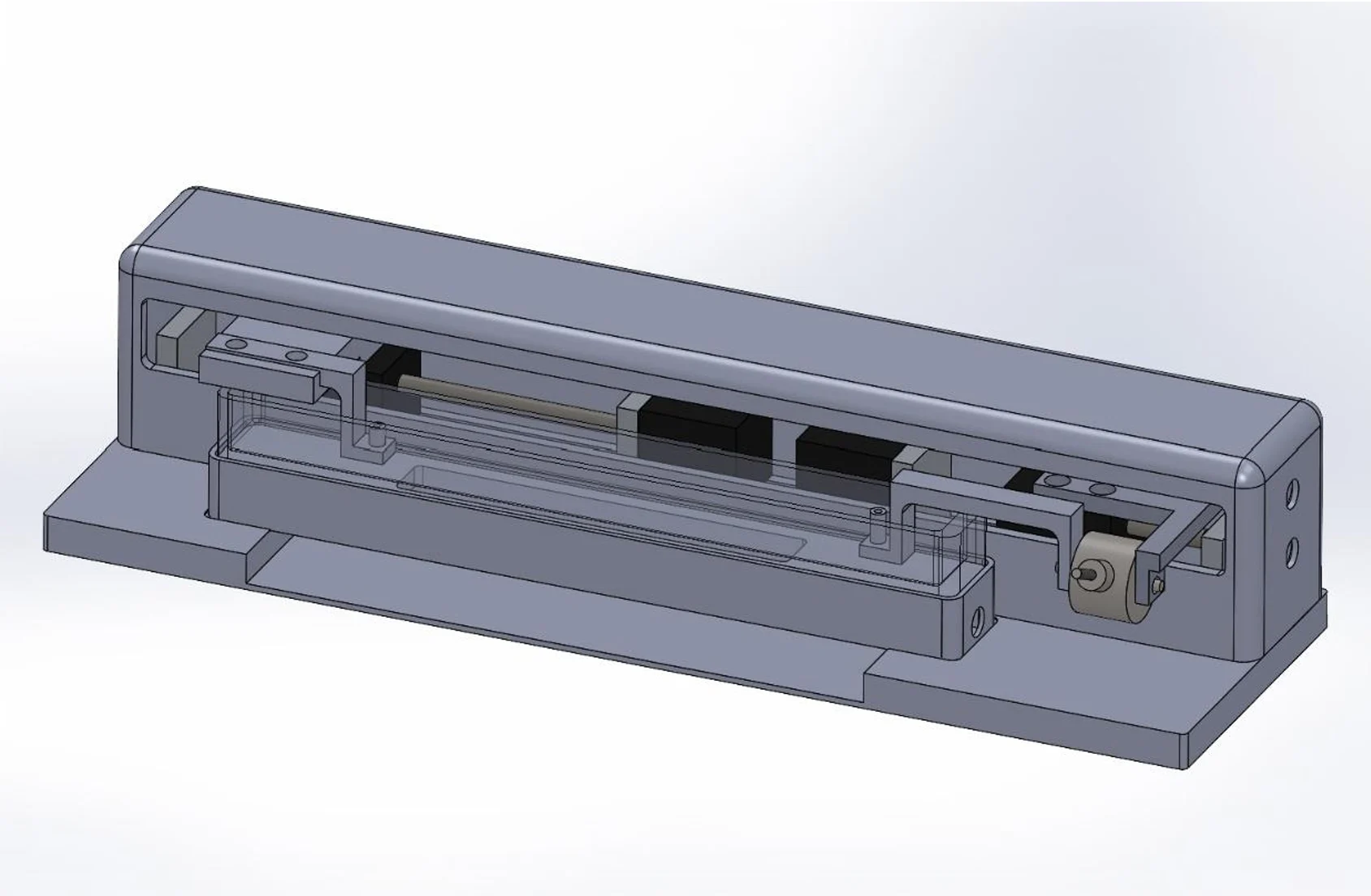
Two linear actuators were incorporated into the specimen-loading assembly, while a third operated a fluid-filled pressure piston.
Axial force was measured using a load cell connected to one specimen mount. Internal pressure was generated by advancing the piston and measured through an electronic pressure sensor connected to the fluid line.
The original engineering proposal specified axial loading up to 10 N and internal pressurization up to 300 mmHg. These design targets were developed around the laboratory’s anticipated specimen sizes and loading requirements.
In the completed software configuration, users could program displacement- or force-controlled axial loading alongside pressure-based inflation protocols, including repeated ramp and sinusoidal loading sequences.


Custom Fixtures for Small Vascular Specimens
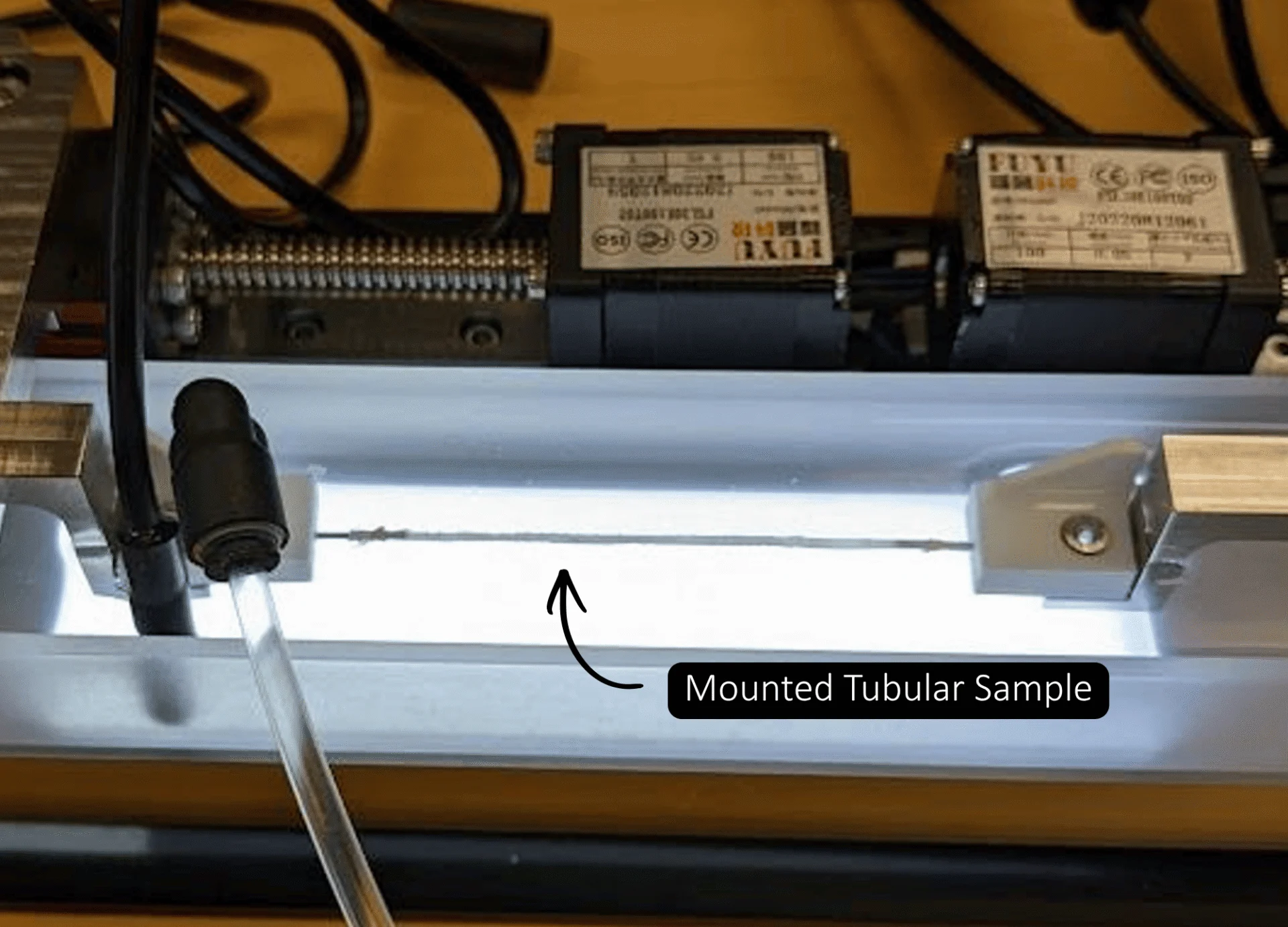
CellScale also developed specimen-mounting arrangements using 3D-printed components and small-diameter stainless steel tubing.
The fixtures allowed tubular samples to be secured at both ends, with one connection supplying pressurizing fluid and the other connected to the axial force measurement assembly.
For particularly small specimens, the mounting instructions describe using stainless steel microtubing as small as 0.5 mm in outer diameter.
This fixture arrangement could be adapted to different specimen sizes while maintaining a sealed connection to the pressure system.
Temperature-Controlled Bath and Optical Diameter Tracking
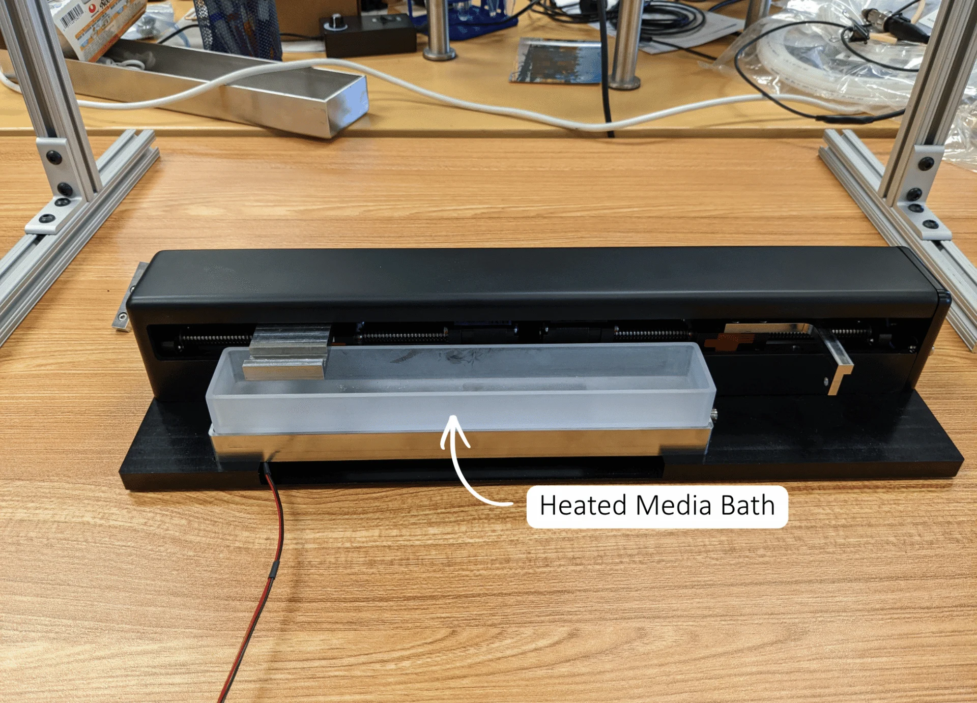
The specimen-loading assembly was positioned within a removable fluid chamber supported by a heated aluminum base. Integrated backlighting improved the contrast between the specimen and its surroundings.
An overhead camera recorded the vessel during testing, while our OpenX software provided image calibration and specimen-edge tracking.
Users can define a tracking region around the tubular specimen, allowing the software to identify upper and lower edges and measure changes in diameter at multiple positions.
The completed system also allows image-based dilation control, where measured specimen expansion can be used as a test control variable.
Together, these measurements allowed researchers to examine how tubular specimens expanded under internal pressure while maintaining defined axial loading conditions.


Results and Scientific Impact
Since the instrument was developed, the Vande Geest laboratory and collaborators have reported its use in three peer-reviewed studies. Although the studies investigate different aspects of vascular graft design, mechanical characterization provides a common starting point.
1. Mechanical Characterization for Hemocompatible Graft Design
In a 2024 study published in the Journal of the Mechanical Behavior of Biomedical Materials, researchers investigated whether a layered vascular graft could generate luminal reversal flows that might reduce platelet deposition.
The custom CellScale system was used to characterize the mechanical response of tubular Tecoflex and thermobonded polycaprolactone constructs during pressure loading.
Experimental measurements were used to fit material parameters for a porohyperelastic computational model. The researchers then examined how different graft constructions affected predicted fluid movement through the graft wall.
In subsequent platelet deposition experiments, the graft design with higher predicted reversal flow showed approximately 75% lower median platelet deposition than the low-reversal-flow design.
Although the CellScale instrument did not measure platelet deposition or fluid reversal directly, its mechanical data contributed to the material model used to investigate the graft designs.
2. Vascular Graft Compliance Testing and Matching to Native Arteries
In research published in Acta Biomaterialia in 2026, the laboratory examined how the initial mechanical compliance of a tissue-engineered vascular graft might relate to subsequent remodeling.
The researchers fabricated multilayered electrospun grafts and adjusted their construction to produce different circumferential mechanical responses.
Using the custom CellScale system, native rat abdominal aortas and engineered grafts were pressurized while changes in outer diameter were recorded.
The measured compliance values were:
- Native rat aorta: 0.0013 ± 0.0004 mmHg⁻¹
- Compliance-matched graft: 0.0011 ± 0.0002 mmHg⁻¹
- Hypocompliant graft: 0.0007 ± 0.00009 mmHg⁻¹
The compliance-matched graft was not significantly different from native aorta under the tested conditions, whereas the hypocompliant design exhibited lower compliance.
This mechanical characterization allowed the researchers to establish distinct graft groups before implantation.

3. Examining Long-Term Graft Remodeling
The mechanically characterized grafts were subsequently implanted into male and female rats and followed over six months.
Although the compliance-matched grafts initially exhibited mechanical behaviour close to that of native aorta, their compliance decreased following implantation. By one month, the initial compliance difference between the engineered graft groups had diminished.
However, other aspects of tissue remodeling remained different.
At six months, the compliance-matched grafts showed greater average material degradation and reduced calcification compared with hypocompliant grafts. The researchers also observed differences in vascular smooth muscle cell phenotype, macrophage presence, collagen deposition, and sex-dependent remodeling.
These findings suggest that initial graft mechanics may influence subsequent biological responses, even when the original compliance difference does not persist throughout implantation.
4. Mechanical Matching in a Longitudinal 3D Culture Model
A separate 2026 study published in Materials Today Bio investigated tissue-graft remodeling using an ex vivo three-dimensional culture platform.
For this work, researchers used a mechanically characterized, compliance-matched graft design as a reference for experiments involving native arterial explants and engineered scaffolds.
The culture platform maintained the cylindrical arrangement of arterial tissue and graft segments, allowing the researchers to repeatedly image tissue-graft interfaces over eight weeks.
Multiphoton imaging was used to follow cellularization and collagen fibre development. The researchers then compared selected features of the cultured grafts with those observed in long-term implanted samples.
Several aspects of collagen organization were similar between the eight-week culture samples and six-month explants, although differences remained between the experimental models.
The custom CellScale device was used for the initial mechanical characterization of the grafts. The subsequent culture and imaging experiments were performed using a separate platform developed by the research team.

Key Engineering Features
Tubular biaxial loading: Independent axial actuation and internal pressurization for small-diameter vascular specimens.
Custom specimen fixtures: Adaptable 3D-printed mounts with stainless steel microtubing for secure, fluid-tight specimen attachment.
Integrated force and pressure measurement: Axial load cell and electronic pressure sensing during mechanical testing.
Temperature-controlled testing: Heated fluid chamber with specimen backlighting for submerged testing.
Optical diameter tracking: Camera-based measurement of specimen dilation at multiple positions along its length.
Programmable OpenX software: Coordinated loading protocols, image acquisition, and specimen deformation tracking.
Related Publications
TITLE
JOURNAL
Journal of the Mechanical Behavior of Biomedical Materials (2024)
APPLICATIONS
RESEARCH SUMMARY
Researchers investigated a layered vascular graft designed to produce luminal reversal flows during cyclic loading. Mechanical characterization of tubular graft materials was performed using a custom CellScale tension and inflation system. The measured responses informed a porohyperelastic computational model used to investigate graft deformation and fluid movement. Subsequent experimental comparisons found lower platelet deposition for the design with higher predicted reversal flow.
Citation: Ali Behrangzade, Sang-Ho Ye, David R. Maestas, William R. Wagner, Jonathan P. Vande Geest, Improving the hemocompatibility of a porohyperelastic layered vascular graft using luminal reversal microflows, Journal of the Mechanical Behavior of Biomedical Materials, Volume 157, 2024, 106638, ISSN 1751-6161, https://doi.org/10.1016/j.jmbbm.2024.106638.
TITLE
JOURNAL
Materials Today Bio (2026)
APPLICATIONS
RESEARCH SUMMARY
A three-dimensional artery-graft culture platform was developed to follow cellularization and collagen remodeling at the interface between native arterial tissue and engineered grafts. A compliance-matched vascular graft design was characterized using the custom CellScale testing system before incorporation into the culture experiments. Longitudinal multiphoton imaging revealed evolving collagen architectures and selected remodeling similarities between cultured grafts and long-term implants.
Citation: David R. Maestas, Trin R. Murphy, Katarina M. Martinet, Tracey Moyston, Leon Xuanyu Min, Ali Behrangzade, Brock J. Pemberton, Jincy Joy, Sang-Ho Ye, George S. Hussey, Mohamad Azhar, William R. Wagner, Jonathan P. Vande Geest, Resolving differential vascular graft remodeling using longitudinal multiphoton tracking in a 3D culture platform, Materials Today Bio, Volume 40, 2026, 103626, ISSN 2590-0064, https://doi.org/10.1016/j.mtbio.2026.103626.
TITLE
JOURNAL
Acta Biomaterialia (2026)
APPLICATIONS
RESEARCH SUMMARY
Researchers used a custom CellScale tubular mechanical testing system to compare the compliance of native rat aortas and multilayered electrospun vascular grafts. The results were used to establish compliance-matched and hypocompliant graft groups before implantation. Six-month follow-up experiments showed differences in graft degradation, calcification, tissue remodeling, and sex-dependent biological responses, even after the initial compliance differences diminished.
Citation: Katarina M. Martinet, David R. Maestas, Leon X. Min, Reyhaneh Gholami, Keishi Kohyama, Jacqueline L. Avila, Sang Ho Ye, Kang Kim, William R. Wagner, Jonathan P. Vande Geest, Long-term remodeling of compliance matched tissue engineered vascular grafts is sex specific, Acta Biomaterialia, Volume 219, 2026, Pages 341-355, ISSN 1742-7061, https://doi.org/10.1016/j.actbio.2026.06.042.
Interested in a Similar Custom Solution?
CellScale works with research laboratories to develop specialized mechanical testing systems when conventional instruments cannot accommodate the required specimen geometry, loading conditions, or measurement methods.
Projects can involve custom actuators, pressure systems, fluid chambers, imaging arrangements, specimen fixtures, and software configurations developed around specific experimental needs.