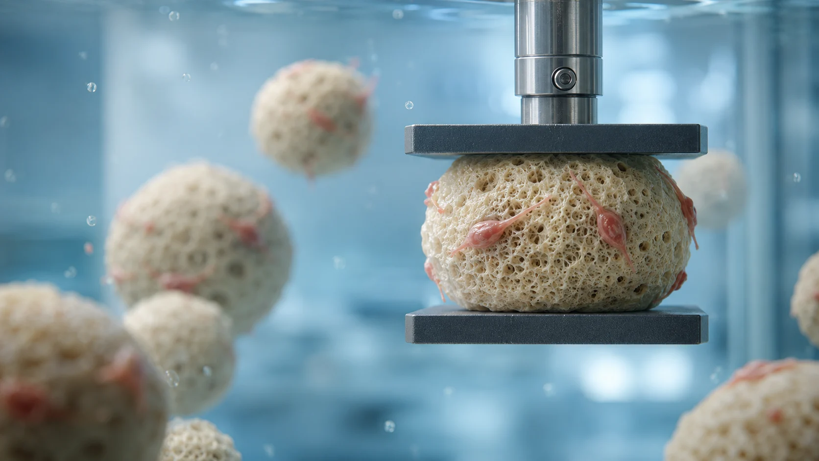



Vascular biomechanics research is essential for understanding how arteries deform, age, remodel, and fail under physiological loading. It is also highly relevant to clinical questions involving peripheral artery disease, diabetes, arterial calcification, stent design, and the effects of limb flexion on vascular mechanics. This collection brings together publications from Dr. Alexey Kamenskiy that used the CellScale BioTester to study these problems through mechanical testing and computational modeling.
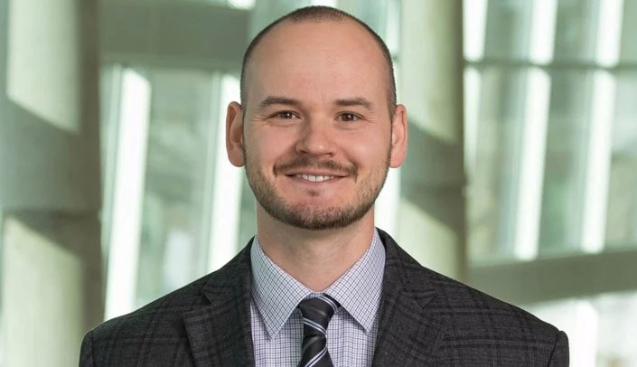
Dr. Alexey Kamenskiy is a professor at the University of Nebraska Omaha and the University of Nebraska Medical Center. His lab focuses on experimental and computational vascular mechanobiology, vascular pathophysiology and aging, and devices and materials for open and endovascular repair. Taken together, the publications below show the breadth of vascular biomechanics research that can be carried out with biaxial tissue testing and related mechanical analysis.
Read more about Vascular Tissue Engineering & Mechanics.
Why this vascular biomechanics research matters
The femoropopliteal artery experiences a particularly complex loading environment. It bends, twists, compresses, and stretches during normal limb motion, while aging, diabetes, and calcification can further alter its mechanical response. Those factors are directly relevant to device durability, arterial injury, and treatment outcomes.
That is why this collection is especially valuable. Rather than focusing on a single experiment, it shows how one research group has built a broader understanding of vascular biomechanics research across constitutive modeling, arterial aging, limb flexion, stiffness, calcification, tissue damage, and stent behaviour.
How the CellScale BioTester supports this work
A common thread across this collection is the CellScale BioTester. The BioTester enables biaxial and soft tissue mechanical testing that supports constitutive modeling, stress-stretch analysis, and the study of vascular tissue behaviour under physiologically meaningful loading conditions.
That makes this page especially useful for readers interested in vascular testing and testing of arteries. The instrument is not used for just one narrow problem. It supports a broader vascular mechanics research program spanning basic science, translational biomechanics, and vascular device design.
Key research themes in this collection
The publications in this collection center on several high-value themes:
Femoropopliteal artery mechanics
Multiple studies focus on how the human femoropopliteal artery deforms under limb flexion and how that behaviour changes with age, disease, and constitutive assumptions.
Arterial aging and diabetes
Several papers examine how aging and diabetes alter arterial stiffening and constitutive response, contributing to a better understanding of vascular remodeling and peripheral artery disease.
Device and stent biomechanics
The collection also includes work comparing femoropopliteal artery stents under axial compression, axial tension, bending, and torsion, which is highly relevant to endovascular repair.
Tissue damage and calcification
Other studies investigate calcification, arterial stiffness, and mechanical damage, linking tissue composition and pathology back to mechanical behaviour.
Computational vascular modeling
Constitutive formulations and modeling studies in this group show how experimentally measured vascular behaviour can be translated into more realistic simulations.
Publications from Dr. Alexey Kamenskiy featuring the CellScale BioTester
Below is the collection of publications currently highlighted from Dr. Kamenskiy’s group:
- The choice of a constitutive formulation for modeling limb flexion-induced deformations and stresses in the human femoropopliteal arteries of different ages
- Constitutive description of human femoropopliteal artery aging
- Constitutive modeling of human femoropopliteal artery biaxial stiffening due to aging and diabetes
- Comparison of femoropopliteal artery stents under axial and radial compression, axial tension, bending, and torsion deformations
- Limb flexion-induced twist and associated intramural stresses in the human femoropopliteal artery
- Prevalence of Calcification in Human Femoropopliteal Arteries and its Association with Demographics, Risk Factors, and Arterial Stiffness
- Cross-sectional pinching in human femoropopliteal arteries due to limb flexion, and stent design optimization for maximum cross-sectional opening and minimum intramural stresses
- Mechanically robust cryogels with injectability and bioprinting supportability for adipose tissue engineering
- Mechanical damage characterization in human femoropopliteal arteries of different ages
- Mechanical stresses associated with flattening of human femoropopliteal artery specimens during planar biaxial testing and their effects on the calculated physiologic stress-stretch state
What this collection shows
What stands out in this collection is the consistency of the application area and the variety of biomechanics questions being addressed. These studies are connected by a common vascular focus, but they span constitutive modeling, device mechanics, arterial pathology, and experimental testing strategy.
That makes this page a strong resource for readers interested in:
- vascular mechanobiology
- femoropopliteal artery biomechanics
- arterial aging biomechanics
- vascular device biomechanics
- computational vascular modeling
Final thoughts
This collection highlights how vascular biomechanics research can build over time when a lab combines careful tissue testing with clinically relevant vascular questions. Dr. Alexey Kamenskiy’s publications show how the CellScale BioTester has supported work on femoropopliteal artery mechanics, arterial aging, diabetes-related stiffening, vascular calcification, stent deformation, and computational modeling.
For readers interested in vascular tissue mechanics, peripheral artery biomechanics, or BioTester-enabled soft tissue testing, this collection is a strong example of how one instrument platform can contribute across an entire research program.
Read more about Dr. Kamenskiy’s research here: UNMC vascular research profile
For related reading, you may also like: