Mechanical Stimulation of Dermal Fibroblasts

CellScale chats with senior researcher Marcel Vlig at the Association of Dutch Burn Centres in Beverwijk, Netherlands. Marcel is studying the cellular response of dermal fibroblasts to mechanical loading using the MechanoCulture B1.

[et_pb_section fb_built=”1″ _builder_version=”4.16″ global_colors_info=”{}”][et_pb_row _builder_version=”4.16″ background_size=”initial” background_position=”top_left” background_repeat=”repeat” global_colors_info=”{}”][et_pb_column type=”4_4″ _builder_version=”4.16″ custom_padding=”|||” global_colors_info=”{}” custom_padding__hover=”|||”][et_pb_text admin_label=”Text” _builder_version=”4.25.2″ global_colors_info=”{}”]

In our In the Lab blog series, we catch up with researchers using CellScale systems.

In this installment of the blog, Caleb Horst chats with senior researcher Marcel Vlig at the Association of Dutch Burn Centres in Beverwijk, Netherlands. Marcel is studying the cellular response of dermal fibroblasts to mechanical loading using the MechanoCulture B1.

Caleb Horst: Thanks Marcel for joining us. I’d like to start just by asking you to tell us a little bit about your research and the goals and what you’re hoping to accomplish.

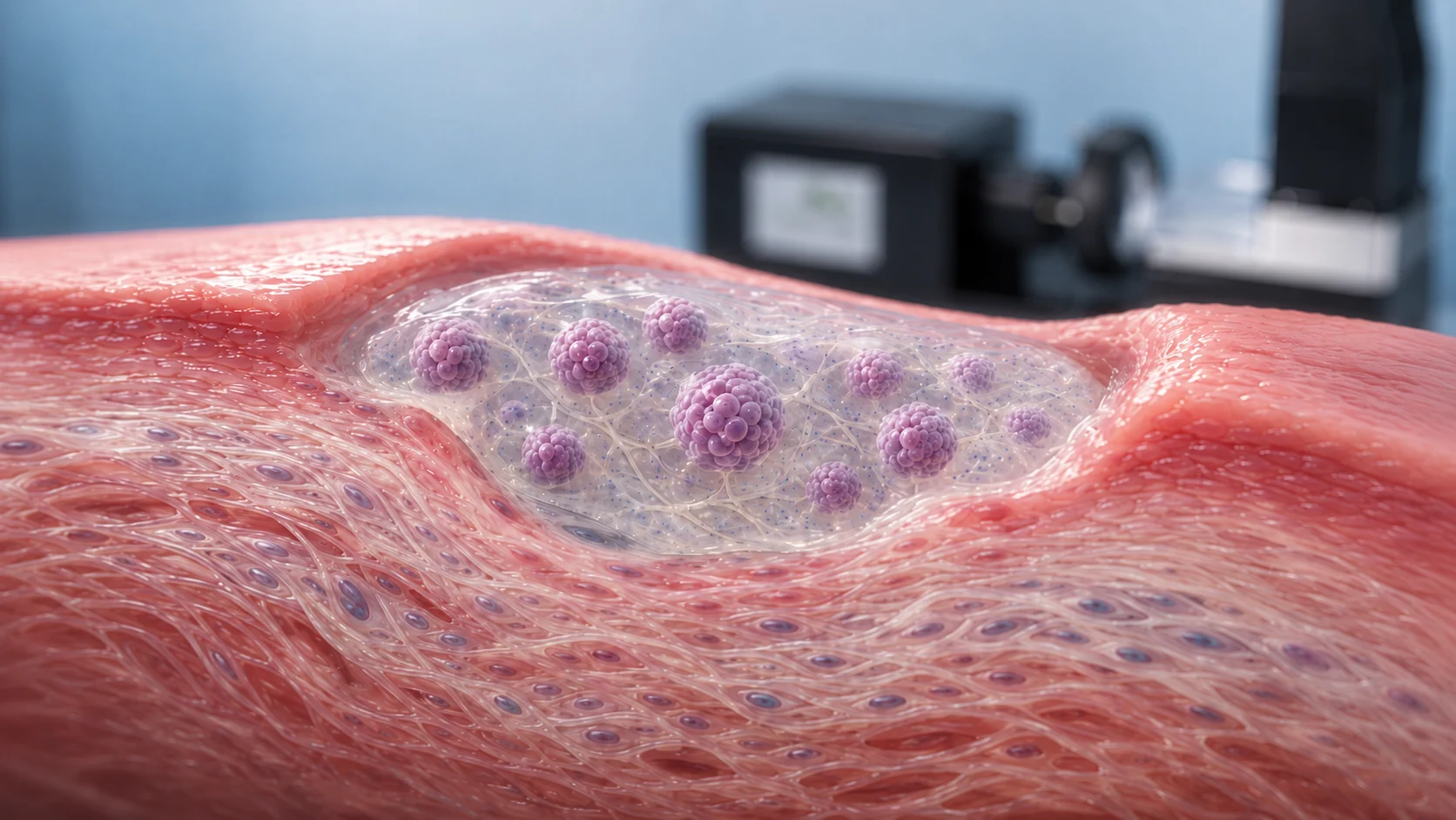
Marcel Vlig: We’re investigating the mechanical tension and mechanical loading to dermal fibroblasts because when people suffer from burn wounds the wounds want to contract. In the clinic I use pins to reverse that contraction and we want to know what the effect is of this mechanical loading to cells.

CH: How are you using the MechanoCulture for the testing that you’re doing, and how does it help you understand the response of the cells?

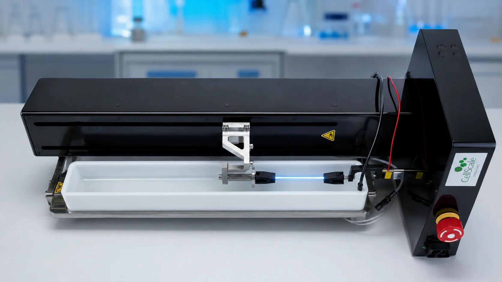
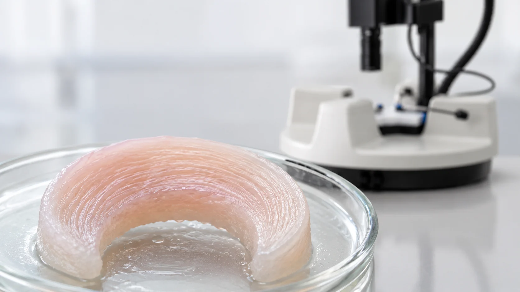
MV: We use the MechanoCulture to set up different stretching regimes; dynamic stretching or static stretching or a combination of these two, to see which one has the most positive effect on dermal fibroblasts. The biggest advantage of the MechanoCulture is that we can use 3D collagen scaffolds to do our experiments with.

CH: Can you walk us through a typical test and just explain to us how you load the cells onto your device and how you run the test?

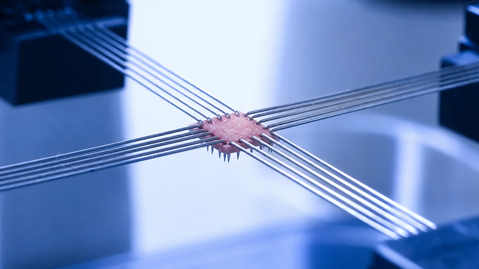
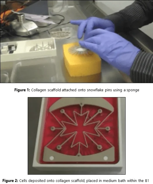
MV: I use a collagen scaffold, which we sterilized previously to our experiment, and then we attach it onto our snowflake using a sponge to push the pins through our collagen scaffold [Figure 1]. We use tweezers to place the collagen scaffold. When it’s in the device we can seed our cells onto the collagen scaffold and we wait for two hours so they can adhere to the scaffold. Then we fill up the chamber with medium, wait for 24 hours so they can expand and grow, and then we start our protocol which is 24 hours of stretch or static stretch [Figure 2], and after that we collect RNA from our cells.

[/et_pb_text][et_pb_image src="https://cellscale.com/wp-content/uploads/2018/10/Blog-4-Fig-1-and-2.png" align="center" align_tablet="center" align_phone="center" align_last_edited="on|desktop" _builder_version="4.16" global_colors_info="{}"][/et_pb_image][et_pb_text _builder_version="4.25.2" global_colors_info="{}"]

CH: And what stage is your research at? Have you been able to show how the cells respond to mechanical loading using the MechanoCulture?

MV: We’ve now established the number of cells we need to seed in our collagen scaffold to obtain proper results and we also see that if we apply a stretch regime on our cells, they do respond to this stress compared to the control cells. So we do have a working system and now we have to apply different kinds of stretch regimes to see which one is more beneficial.

CH: Thanks Marcel, and thank you for watching this video. If you have any questions about CellScale’s products please feel free to contact us.


Watch the interview online: https://www.youtube.com/watch?v=gsknXqPFDQM

CTA: Learn more about CellScale’s MechanoCulture B1 or check out some of our customers’ publications.

[/et_pb_text][/et_pb_column][/et_pb_row][/et_pb_section]

CELLSCALE INSTRUMENT USED

TAGS

Association of Dutch Burn Centres, Burn Injury Research, Cellular Mechanobiology, Collagen Scaffolds, Dermal Fibroblasts, Mechanical Stimulation, MechanoCulture B1, Wound Healing Research

POST DETAILS
CATEGORY

Research Highlights

INSTRUMENT USED
RESEARCH APPLICATIONS

Related Posts:

Filter by Category:
Filter by Instrument
Filter by Testing Method:
Filter by Research Application:
MSC spheroids embedded in a translucent PEG-fibrin hydrogel within soft vocal fold-like tissue, with a blurred CellScale MicroTester in the background.

June 11, 2026

Vocal Fold Regeneration with MSC Spheroids

Hydrogel eye drop on a corneal surface with a mechanical testing probe and force-distance curve representing mucoadhesion testing for ocular drug delivery.

May 28, 2026

Mucoadhesion Testing of Hydrogel Eye Drops for Ocular Drug Delivery

May 14, 2026

Flexible Flow Sensor Testing for Pioneering Autonomous Robotics

Featured illustration showing a layered tissue phantom under compression, an elastography heatmap, a phase velocity versus depth graph, and an AI neural network used to support depth-resolved stiffness analysis.

May 7, 2026

Validating AI-Based Elastography Measurements with Compression Testing

Illustration of MSC hydrogel microspheres for diabetic wound healing placed in a wound bed with inflammation on the left and tissue repair on the right

April 30, 2026

MSC Hydrogel Microspheres for Diabetic Wound Healing

A porcine pericardium sample on a white bench, with arrows indicating biaxial mechanical testing directions

April 23, 2026

Porcine Pericardium Biaxial Testing for Heart Valve Design

Contact Sales

Product of Interest: