IVD Regenerative Medicine featuring Pullulan Microbeads in an Injectable System

A tensile test divider icon

Dr Catherine Le Visage from the Université de Nantes in France shares part of her major research into regenerative medicine to treat intervertebral disc degeneration disease. Her group at INSERM created an injectable system that deliver growth factors through the use of microbeads and tested the system in vitro.

Top left: Pullulan microbeads observed with a confocal microscopy Bottom left: Pullulan microbeads observed with a scanning electron microscope Right: Pullulan microbeads mechanical characterization. A hydrated single PMB was compressed between 2 plates using a MicroTester. Force and displacement data were recorded and a representative curve was presented.
CellScale hexagon shapes

Dr Catherine Le Visage and her group at the Regenerative Medicine and Skeleton (INSERM U119) based in the School of Dental Surgery, University of Nantes – France has derived an ingenious way to treat discogenic low back pain, something several of us experience as we age.

A common reason of low back pain is the degeneration of the intervertebral disc (IVD), which is a fibrocartilaginous tissue located between each vertebrae in our spine. Current treatment does not address the etiological cause of disc degenerative disease (DDD) thus regenerative medicine approaches are considered with deep interest – in particular, the stimulation of local cells by in situ injection of growth factors targeting IVD degenerative process.

Dr Le Visage’s approach is an injectable biphasic system of pullulan microbeads (PMBs) dispersed within a cellulose-based hydrogel (Si-HPMC). Pullulan is a neutral, linear and non-immunogenic polysaccharide that is widely used because of its natural properties (biodegradable, biocompatible, non-toxic). The research team demonstrated a sustained release of growth factors TGF-β1and GDF-5 in vitro for up to 28 days, while maintaining biological activity on human cells. In addition, they were able to measure for the first time the mechanical properties of the PMBs through a parallel plate compression test with the CellScale MicroTester.
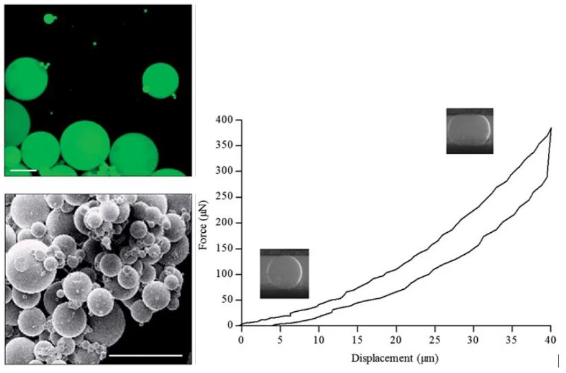
Top left: Pullulan microbeads observed with a confocal microscopy Bottom left: Pullulan microbeads observed with a scanning electron microscope Right: Pullulan microbeads mechanical characterization. A hydrated single PMB was compressed between 2 plates using a MicroTester. Force and displacement data were recorded and a representative curve was presented.
Top left: Pullulan microbeads observed with a confocal microscopy
Bottom left: Pullulan microbeads observed with a scanning electron microscope
Right: Pullulan microbeads mechanical characterization. A hydrated single PMB was compressed between 2 plates using a MicroTester. Force and displacement data were recorded and a representative curve was presented.

This study is part of a larger comprehensive and continuous study to improve treatment options of DDD through regenerative medicine.

You can read more about Dr Le Visage’s work here.

Read the full journal publication here.

To read about a collagenous bioscaffold for the treatment of cutaneous wounds, click here.

CellScale hexagons, without text

CELLSCALE INSTRUMENT USED

TAGS

compression testing, Disc degeneration, Growth factor delivery, Injectable biomaterials, Intervertebral disc research, MicroTester, Pullulan microbeads, regenerative medicine, Spine Biomechanics

POST DETAILS
CATEGORY

Research Highlights

INSTRUMENT USED
MicroTester
RESEARCH APPLICATIONS
Drug Screening & Drug Delivery MechanicsInjectable & Regenerative BiomaterialsIntervertebral Disc Biomechanics
TESTING METHODS
Compression TestingHydrated and Temperature Controlled TestingMicro-Mechanical Testing

Related Posts:

Filter by Category:
Filter by Instrument
Filter by Testing Method:
Filter by Research Application:
UniVert performing hydrated tensile testing on a glowing hydrogel optical fibre mounted between grips in a fluid bath.

July 16, 2026

Hydrogel Optical Fiber Testing Under Repeated Strain

Bioprinted meniscus tissue engineering construct with curved collagen-like alignment patterns in the foreground and a softly blurred CellScale BioTester in the background.

July 2, 2026

Meniscus Tissue Engineering with Boundary-Guided Collagen Alignment

Hydrogel fiber mechanical testing setup showing a thin hydrated fibre suspended across two supports while a small MicroTester beam presses downward in the centre, before and after.

June 25, 2026

Hydrogel Fibre Mechanical Testing of Recombinant Hagfish Protein Materials

MSC spheroids embedded in a translucent PEG-fibrin hydrogel within soft vocal fold-like tissue, with a blurred CellScale MicroTester in the background.

June 11, 2026

Vocal Fold Regeneration with MSC Spheroids

Hydrogel eye drop on a corneal surface with a mechanical testing probe and force-distance curve representing mucoadhesion testing for ocular drug delivery.

May 28, 2026

Mucoadhesion Testing of Hydrogel Eye Drops for Ocular Drug Delivery

May 14, 2026

Flexible Flow Sensor Testing for Pioneering Autonomous Robotics

Contact Sales

Product of Interest:
CellScale hexagon shapes