Skeletal muscle is mechanically active tissue. Its cells develop in an environment where stiffness, structure, movement, and electrical activity are all present at the same time. Trying to reproduce even a few of those cues in a tissue-engineered scaffold can get complicated fairly quickly.
A 2026 study in ACS Applied Materials & Interfaces approached that problem by developing a piezoelectric hydrogel for skeletal muscle tissue engineering that combined muscle-relevant stiffness, 3D-printed microchannels, and an electrical response triggered by ultrasound.
Researchers from Cleveland State University built the platform from gelatin methacrylate (GelMA), poly(acrylic acid) (PAA), and barium titanate (BTO) nanoparticles. They used a CellScale UniVert to measure the hydrogel platforms in compression while working toward mechanical properties relevant to skeletal muscle.
The study was motivated by volumetric muscle loss, or VML, where enough skeletal muscle is lost that normal regeneration is severely limited. This was not a VML treatment study, though. The work stayed in vitro and focused on how different material and stimulation cues affected C2C12 myoblast development.
Why a Piezoelectric Hydrogel for Skeletal Muscle Tissue Engineering?
A scaffold intended for skeletal muscle tissue engineering has a few different jobs to do. It needs to support cells physically, but the surrounding mechanics can also influence how those cells attach, spread, differentiate, and organize.
The researchers wanted their platform to add something else: bioelectrical cues.
Piezoelectric materials generate an electrical response when they are mechanically deformed. In this study, the idea was to use ultrasound to deform the material without wires or implanted electrodes. That makes the platform part of a broader class of stimuli-responsive hydrogels where an external stimulus changes the material response.
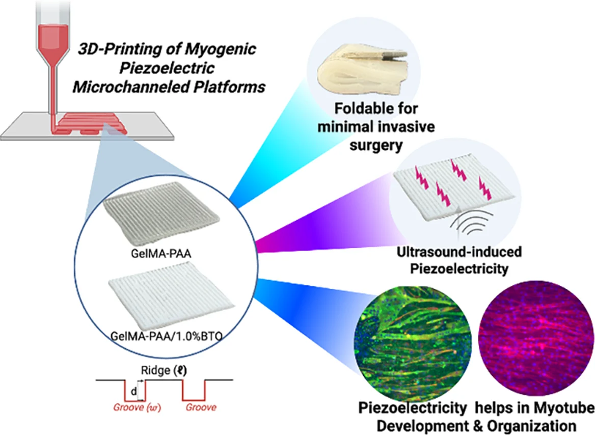
For this piezoelectric hydrogel for skeletal muscle tissue engineering, BTO nanoparticles were incorporated into a GelMA-PAA network. The hydrogel was then extrusion printed with aligned microchannels. Those channels were intended to provide physical guidance, while stiffness supplied a mechanical cue and ultrasound-induced electroactivity added another layer.
Before getting to the cell work, however, the researchers first had to decide what the hydrogel itself should feel like mechanically.
Tuning Hydrogel Stiffness Before 3D Printing
The amount of GelMA had a fairly clear effect on the compression behaviour of the material.
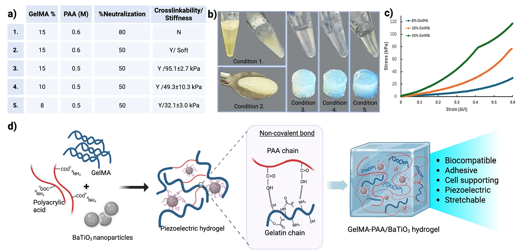
With PAA concentration and neutralization held constant, the formulation containing 15% GelMA had a reported stiffness of about 95.1 kPa. Reducing GelMA to 10% brought that value to about 49.3 kPa. At 8% GelMA, stiffness fell to roughly 32.1 kPa.
The authors selected the 8% GelMA formulation because they considered it closer to native skeletal muscle. This is a useful example of why hydrogel mechanical testing can become part of material development rather than simply a final characterization step.
The material formulation was being changed, the mechanical response was measured, and those measurements helped determine which formulation moved forward.
Similar questions come up regularly with GelMA-based materials. We have looked previously at how material formulation and mechanics interact in GelMA bioinks used for 3D bioprinting, where printability and mechanical behaviour also have to be considered together.
For a piezoelectric hydrogel for skeletal muscle tissue engineering, the situation gets another layer of complexity because adding the electroactive component can change the mechanics too.
How the UniVert Was Used to Measure Scaffold Stiffness
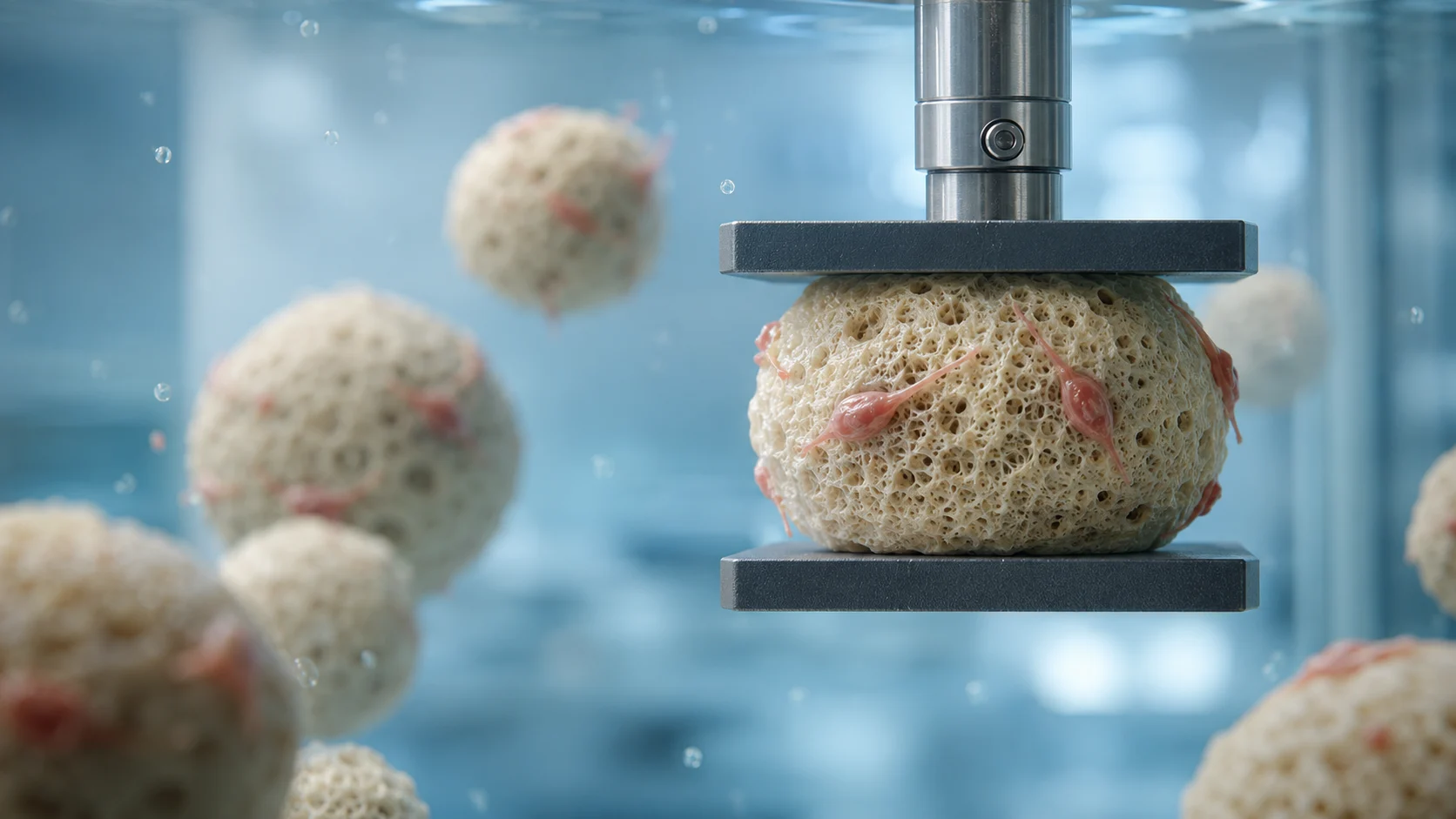
Once the platforms had been fabricated, the researchers used the UniVert for uniaxial compression testing.
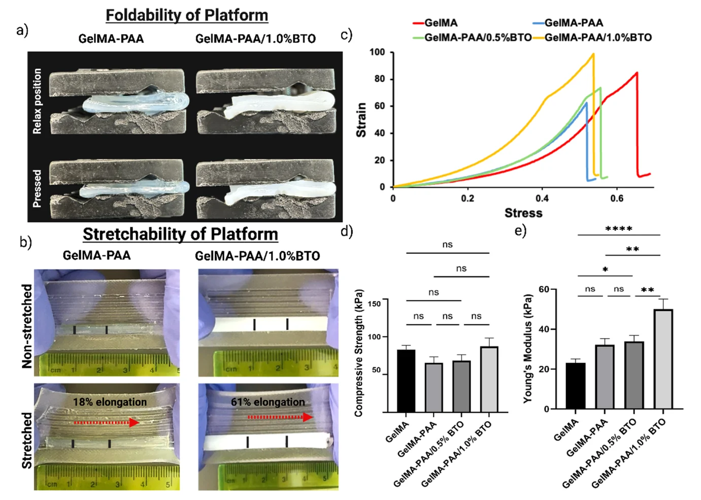
Cylindrical specimens measuring 9 mm in diameter and 4 mm thick were placed between 25 mm compression platens. Testing was performed at room temperature with a 10 N load cell and a crosshead speed of 1 mm/min. Force-displacement and stress-strain curves were recorded, and Young’s modulus was calculated from the stress-strain response.
This is the part of the study where the piezoelectric hydrogel for skeletal muscle tissue engineering connects most directly to scaffold mechanical testing.
Adding BTO changed the response. The GelMA-PAA/1.0% BTO platform had the highest reported compressive strength, about 92.6 ± 8.7 kPa, although the differences in compressive strength among formulations were not statistically significant. The effect on Young’s modulus was clearer. Stiffness increased as PAA and BTO were incorporated, reaching roughly 50 kPa for the 1.0% BTO formulation.
That value remained within the range the authors were targeting for muscle-like mechanics.
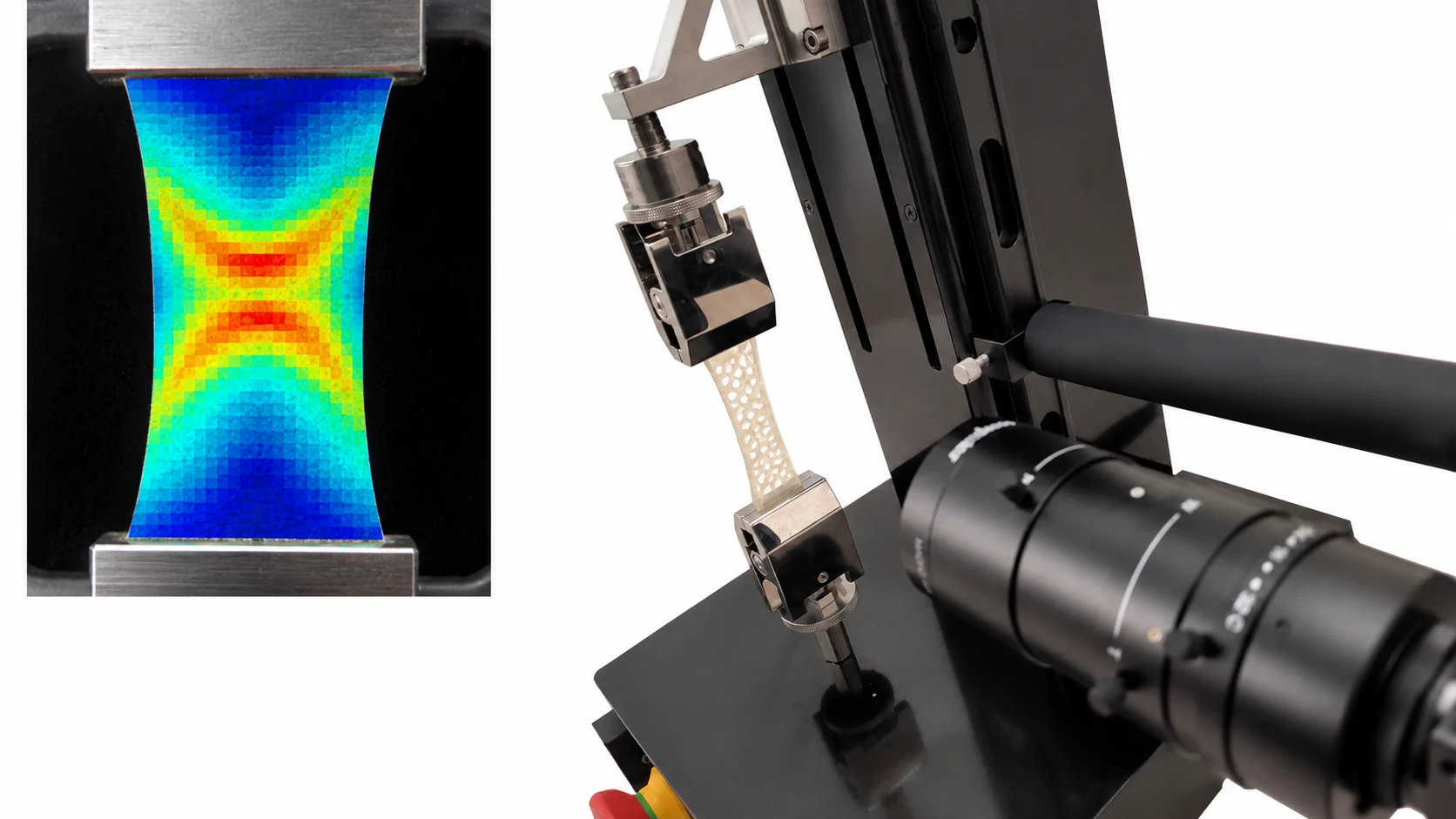
One thing worth keeping separate is the stretchability shown in the figure below. The photographs in panel (b) came from a separate manual stretch assessment, not tensile testing on the UniVert. BTO incorporation increased the reported elongation from 18% to 61%, while the UniVert measurements in panels (c) through (e) deal with compression behaviour.
For more on why methods and analysis choices matter when reporting soft-material stiffness, see our earlier article on hydrogel stiffness measurement.
Ultrasound Activation of a Piezoelectric Hydrogel for Skeletal Muscle Tissue Engineering
The next question was whether mechanical deformation from ultrasound could produce a useful electrical response.
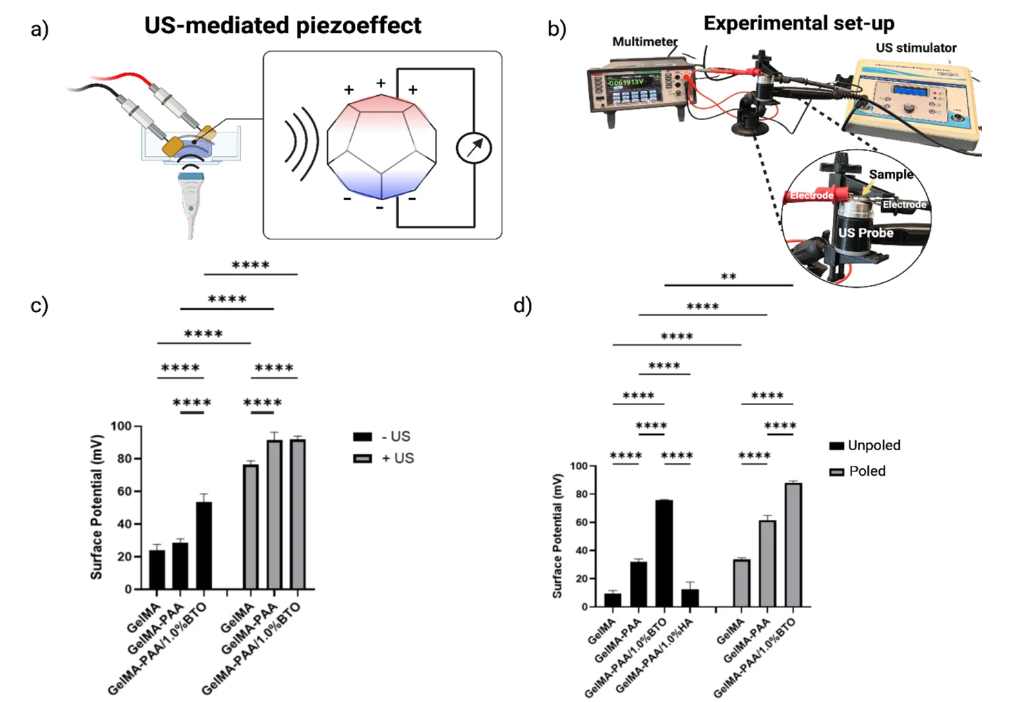
The researchers positioned hydrated or lyophilized hydrogel samples between copper electrodes and applied continuous ultrasound at 1 MHz and 0.5 W/cm². Surface potential was then measured under open-circuit conditions.
The hydrated results need a little care in how they are interpreted.
Under ultrasound, both GelMA-PAA and GelMA-PAA/BTO approached surface potentials of about 90 mV. In other words, the hydrated electrical response could not simply be attributed to BTO. The authors suggest that PAA contributes its own hydration-dependent electroactivity as ions and interfacial charges move within the water-rich polymer network.
They therefore repeated parts of the experiment using lyophilized samples.
Without the water-rich environment, GelMA-PAA produced a considerably smaller response. The BTO-containing material remained much more electroactive, reaching about 75 mV in the unpoled condition and close to 90 mV after poling. A hydroxyapatite nanoparticle control did not show the same increase.
So the piezoelectric hydrogel for skeletal muscle tissue engineering was really a combined electroactive system. Hydrated PAA contributed to the electrical response, while BTO added a distinct solid-state piezoelectric component.
That distinction matters later when interpreting what happened to the cells. Ultrasound was not only producing an electrical signal. It was mechanically stimulating the scaffold as well.
How Microchannels Affected Myotube Alignment
The printed architecture added another variable.
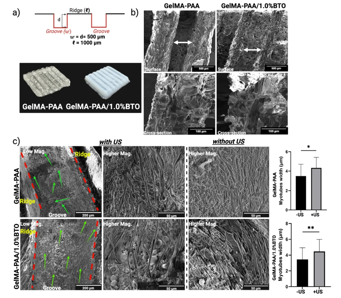
The upper surface of each scaffold contained parallel ridges and grooves, with 1 mm ridges separated by 0.5 mm grooves. C2C12 myoblasts were cultured on these microchanneled platforms and differentiated toward myotubes.
By day 7, the SEM images showed myotubes following the longitudinal direction of the printed ridges.
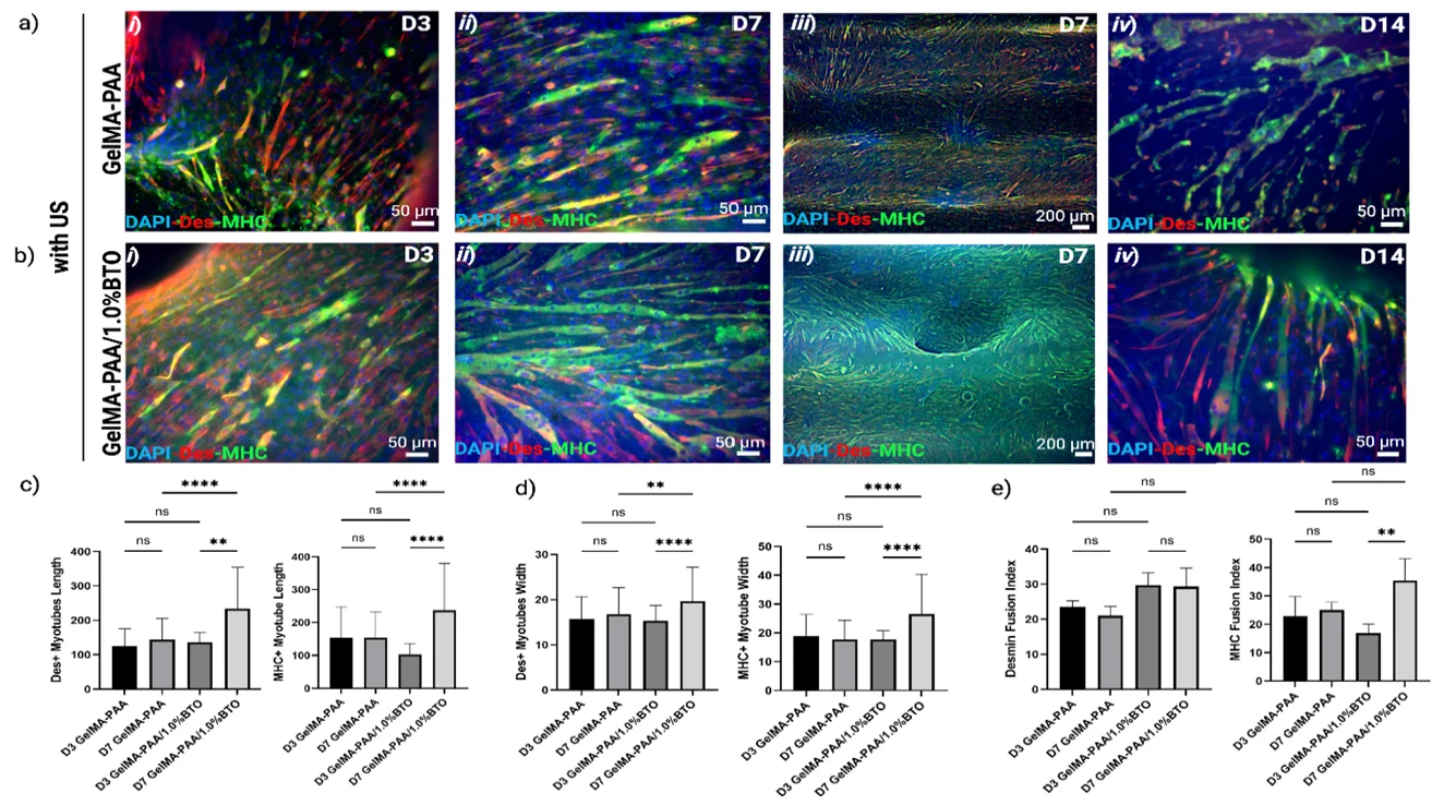
This is one of the more visually convincing parts of the study. The physical channels appear to have provided the main directional cue. Ultrasound then increased myotube width on both GelMA-PAA and GelMA-PAA/BTO platforms, while organization was more pronounced on the BTO-containing material under ultrasound.
That means the different parts of the piezoelectric hydrogel for skeletal muscle tissue engineering were not all doing the same job.
The microchannels guided direction. Ultrasound supplied mechanical stimulation. BTO was associated with changes in myotube growth and organization and, when mechanically activated, contributed the additional piezoelectric response.
Immunofluorescence measurements supported a similar picture. Myosin heavy chain positive myotubes were wider on the BTO-containing platforms, and F-actin appeared more elongated and parallel. The strongest organization was generally observed when BTO-containing platforms were exposed to ultrasound.
The authors also measured intracellular calcium fluorescence, but they are appropriately cautious about that result. These were static fluorescence measurements, not dynamic calcium transients or contractility tests, so they do not demonstrate that the resulting myotubes were functionally contracting.
Stimulation Timing Mattered
A useful part of this study is that the researchers did not stop at a single day-7 endpoint.
They followed myogenic development over time while continuing ultrasound treatment. At day 3, differences between the platforms were still fairly limited. Some early myogenic markers had changed, but there were not yet clear differences in myotube length, width, or fusion across most measurements.
By day 7, the picture had changed.
On the BTO-containing piezoelectric hydrogel for skeletal muscle tissue engineering, desmin-positive and MHC-positive myotubes were longer and wider than at day 3, and several measures of maturation had increased. MYH2 expression was also elevated at day 7.
The mechanotransduction data moved on a similar timescale. FAK and ERK phosphorylation were not significantly different after ultrasound at day 3, but both were higher in the ultrasound-treated BTO group by day 7. It appears that the cells were responding to an evolving electromechanical environment rather than producing an immediate, fixed response.
Then came day 14.
Continued daily ultrasound did not produce another step forward. Instead, the researchers observed loss of myotube structural integrity, reduced desmin staining, and disrupted architecture.
That result makes the study more useful. A piezoelectric hydrogel for skeletal muscle tissue engineering may provide several potentially helpful cues, but the dose and timing of those cues still matter. Under the conditions tested here, the first seven days appeared more favourable than continued daily stimulation through day 14.
Where This Leaves Skeletal Muscle Tissue Engineering
There is an appealing idea behind this work: instead of asking one material property to control cell behaviour, build several cues into the same scaffold and then look at how they interact.
The piezoelectric hydrogel for skeletal muscle tissue engineering combined mechanics, topography, ultrasound stimulation, and electroactivity. In practice, though, those variables are difficult to separate perfectly. Ultrasound mechanically stimulates the scaffold even without BTO. PAA itself contributes to electrical behaviour when hydrated. BTO changes stiffness as well as electroactivity.
That is why the control groups in this study matter as much as the final BTO-plus-ultrasound condition.
It is also worth keeping the scale of the work in mind. These were C2C12 cells in vitro. The study did not test muscle force production, functional contraction, implantation, vascularization, innervation, or repair of a volumetric muscle loss defect. The authors identify functional characterization and in vivo testing as next steps.
The question of what counts as “muscle-like stiffness” is not particularly simple either. Real skeletal muscle mechanics and regional stiffness depend on tissue orientation, anatomical location, loading state, and measurement method. A single Young’s modulus does not capture all of that.
Still, tuning scaffold mechanics before evaluating the biological response gives the experiment a useful foundation. For a piezoelectric hydrogel for skeletal muscle tissue engineering, stiffness was not just another value added to the characterization table. It was one of the properties the researchers actively adjusted as they developed the material.
Citation
Derya Ozhava, Surendrasingh Y Sonaye, Breanne L Welsh, Phuong T Nguyen, Yavar Shiravand, Kara E Timinski, Karim Elhattab, Keith Coasey, Kailash Gulshan, Prabaha Sikder; Flexible 3D-Printed Piezoelectric and Bioinstructive Platforms for Skeletal Muscle Development and Organization. ACS Appl. Mater. Interfaces 2026; https://doi.org/10.1021/acsami.6c11224
Using the UniVert for Hydrogel and Scaffold Compression Testing
In this study, the UniVert was used in a fairly straightforward way: cylindrical hydrogel platforms were positioned between compression platens, loaded at a controlled rate, and the resulting force and displacement data were used to generate stress-strain curves and calculate Young’s modulus.
That same approach is commonly useful when comparing hydrogel formulations, tissue-engineering scaffolds, and other compliant materials where changes in composition can affect stiffness and load response.
The UniVert is a uniaxial mechanical tester used for controlled tensile and compression testing of biological tissues, biomaterials, hydrogels, scaffolds, polymers, and other soft materials. In a compression setup, researchers can use platens and an appropriately selected load cell to apply controlled displacement while recording force throughout the test.
For work on a piezoelectric hydrogel for skeletal muscle tissue engineering, that mechanical characterization can sit alongside cell culture, imaging, rheology, or other assays rather than being treated as a separate experiment. Here, it helped the researchers compare formulations and check whether the final material remained within the stiffness range they wanted before looking at myotube development.