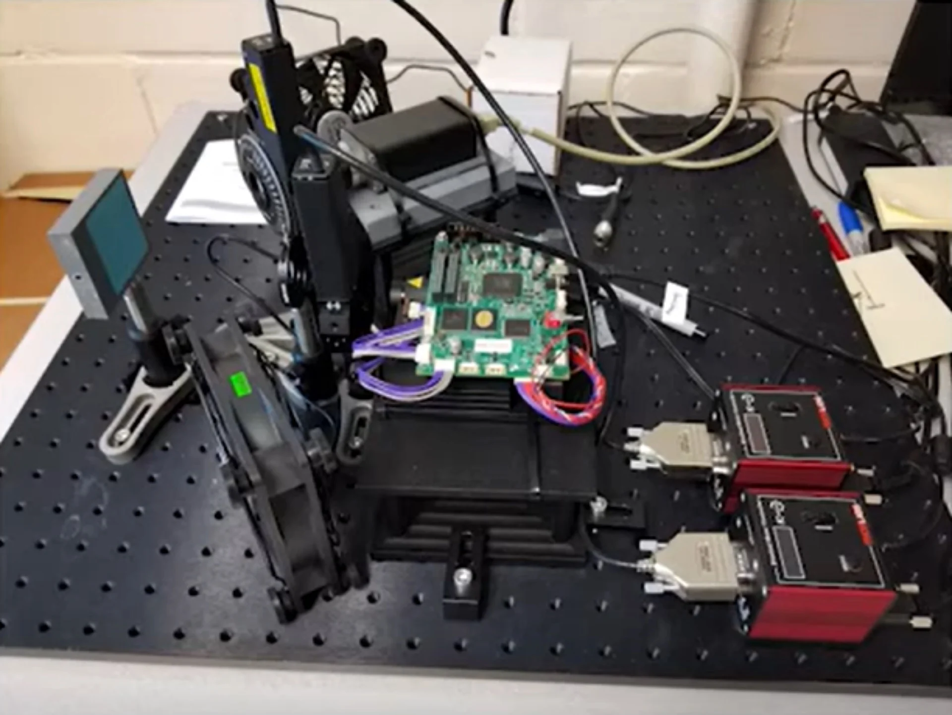
Heart Valve Biaxial Testing with Integrated Imaging

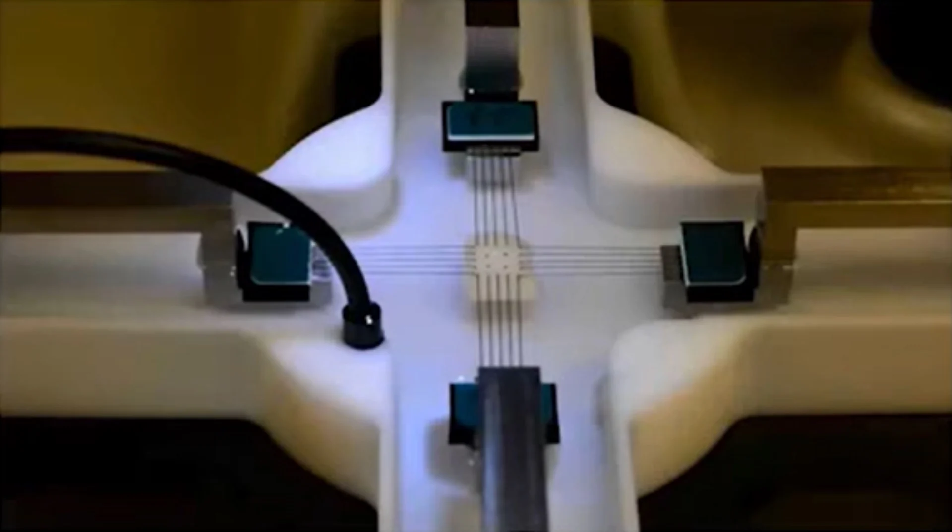


University of Oklahoma
Biomechanics and Biomaterials Design Laboratory with Dr. Chung-Hao Lee
Project Background
Accurate heart valve constitutive modeling requires high-quality experimental data describing both macroscopic mechanical behaviour and underlying tissue microstructure. While biaxial mechanical testing provides essential stress–strain information, traditional approaches offer limited insight into how collagen fiber architecture evolves during deformation.
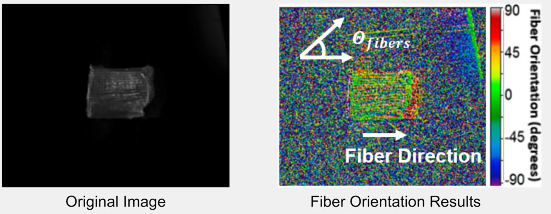
Professor Chung-Hao Lee’s group at the University of Oklahoma sought to bridge this gap by coupling heart valve biaxial testing with real-time optical imaging. Their objective was to directly observe collagen fiber reorientation during loading and integrate these data into advanced valve constitutive modeling and finite element models of heart valve tissue.
CellScale’s BioTester provided a robust and customizable biaxial heart valve mechanics testing platform that was adapted to support the laboratory’s novel imaging approach.

Dr. Chung-Hao Lee
University of Oklahoma


The Challenge
Linking Mechanics and Microstructure
Heart valve function is governed by the interaction between tissue-level mechanics and collagen fiber organization. Conventional mechanical tests measure forces and displacements but cannot directly resolve microstructural changes occurring during loading.
Real-Time Imaging During Heart Valve Biaxial Testing
- Operating concurrently with biaxial mechanical tests
- Capturing spatially resolved collagen orientation data
- Maintaining optical access without interfering with mechanical loading
Integration with Constitutive Modeling
Experimental data needed to support calibration and validation of advanced hyperelastic material models used in finite element simulations of biaxial heart valve mechanics.
Custom Solution Developed by CellScale
Biaxial Mechanical Testing Platform
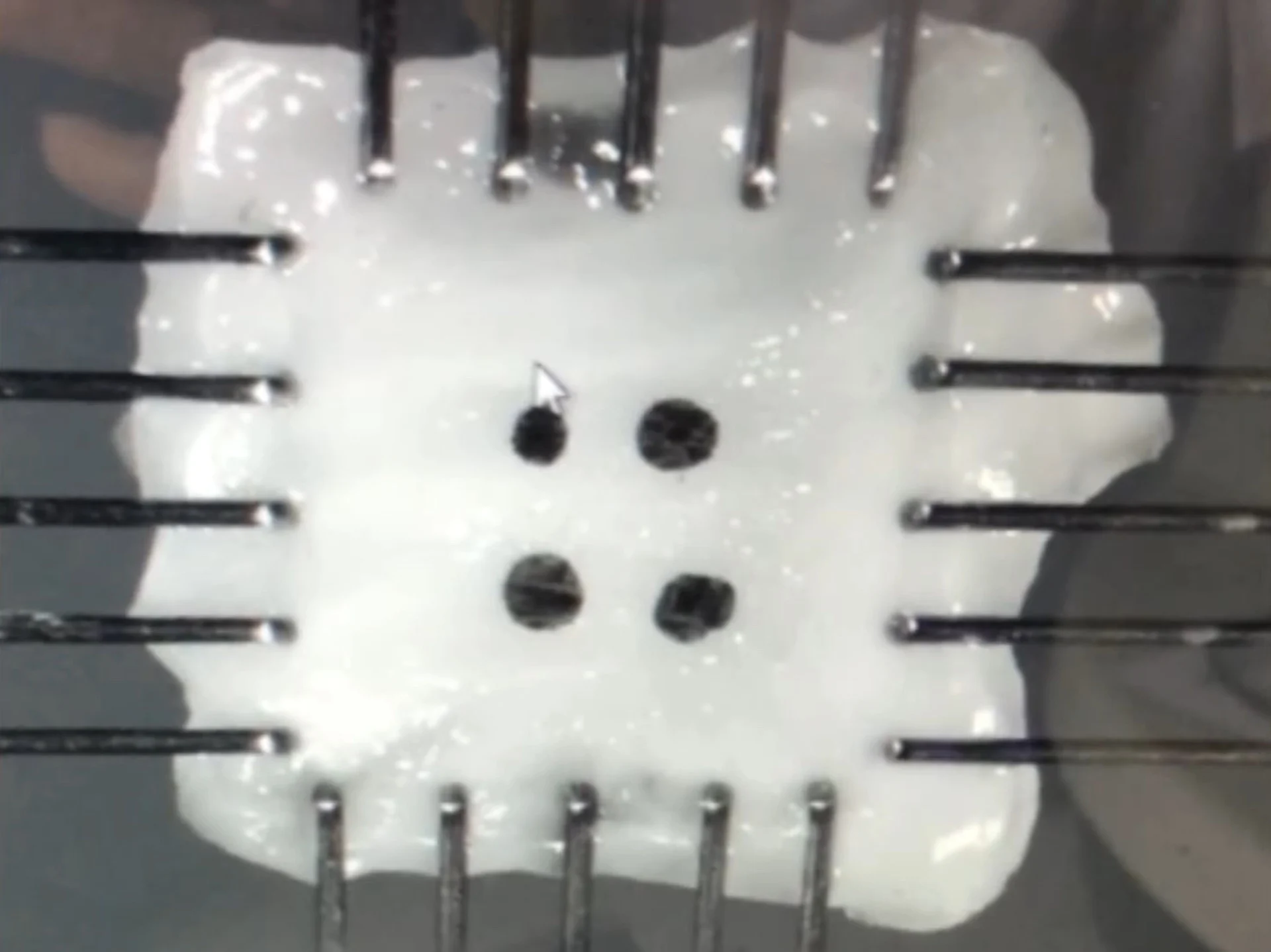
The CellScale BioTester enabled precise equibiaxial and non-equibiaxial loading of heart valve tissue samples under physiological conditions.
Key features included:
- Independent force and displacement control in two orthogonal axes
- Submerged testing capability for hydrated soft tissues
- High-resolution force and displacement measurement
- Compatibility with BioRakes and alternative mounting strategies
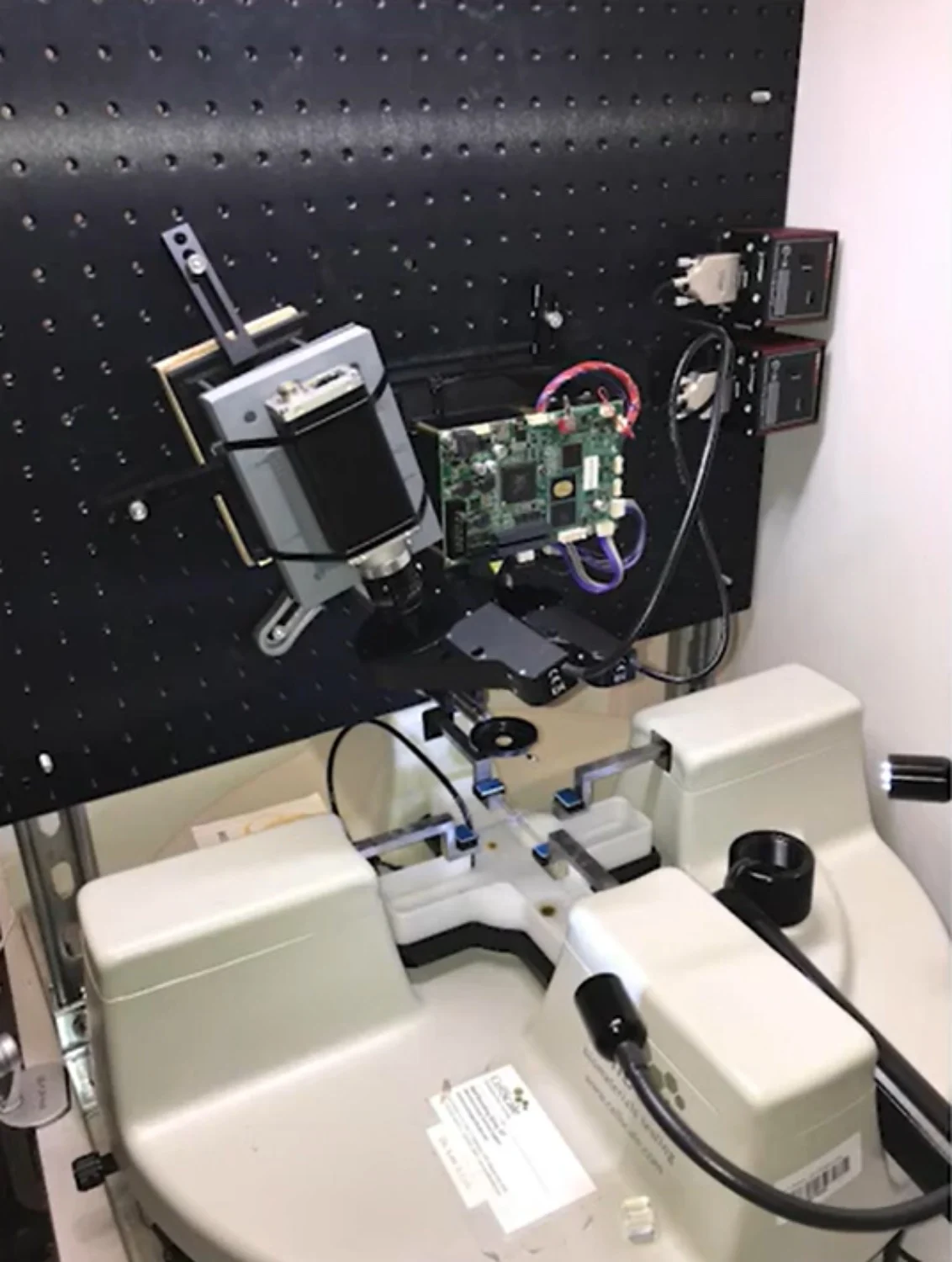
Integrated Imaging Mechanical Testing System
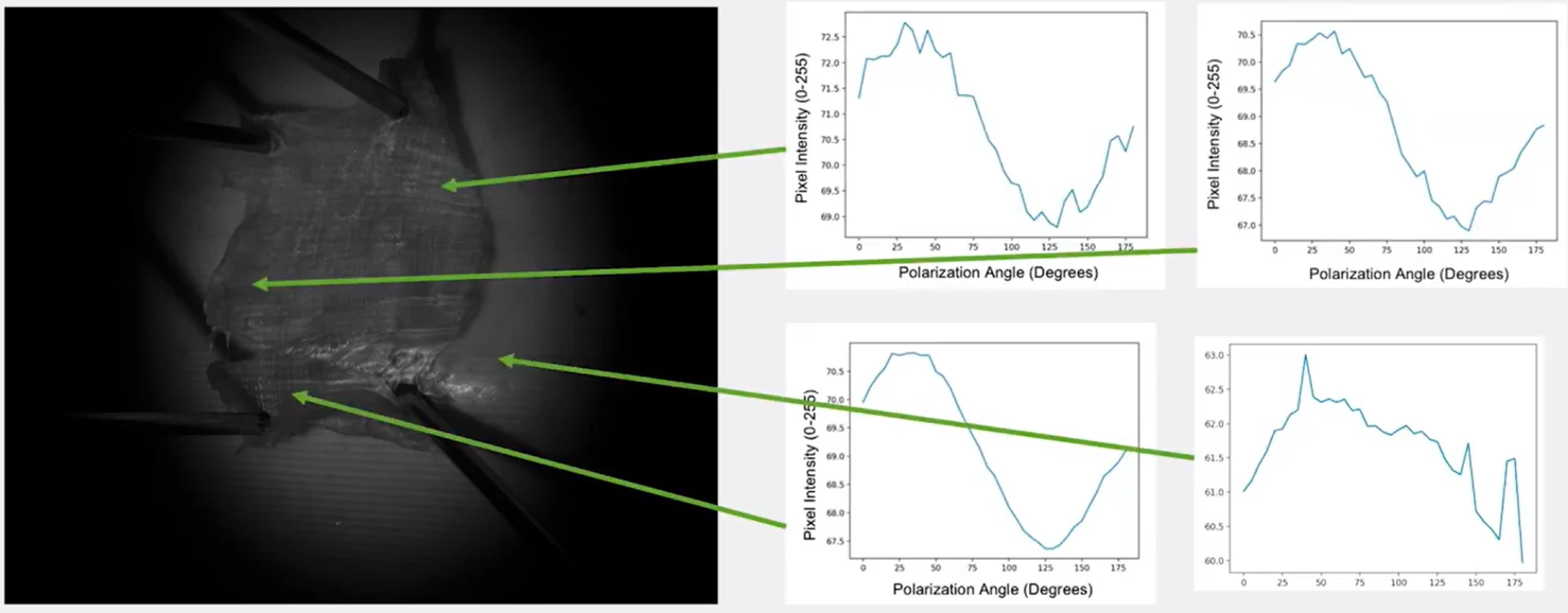
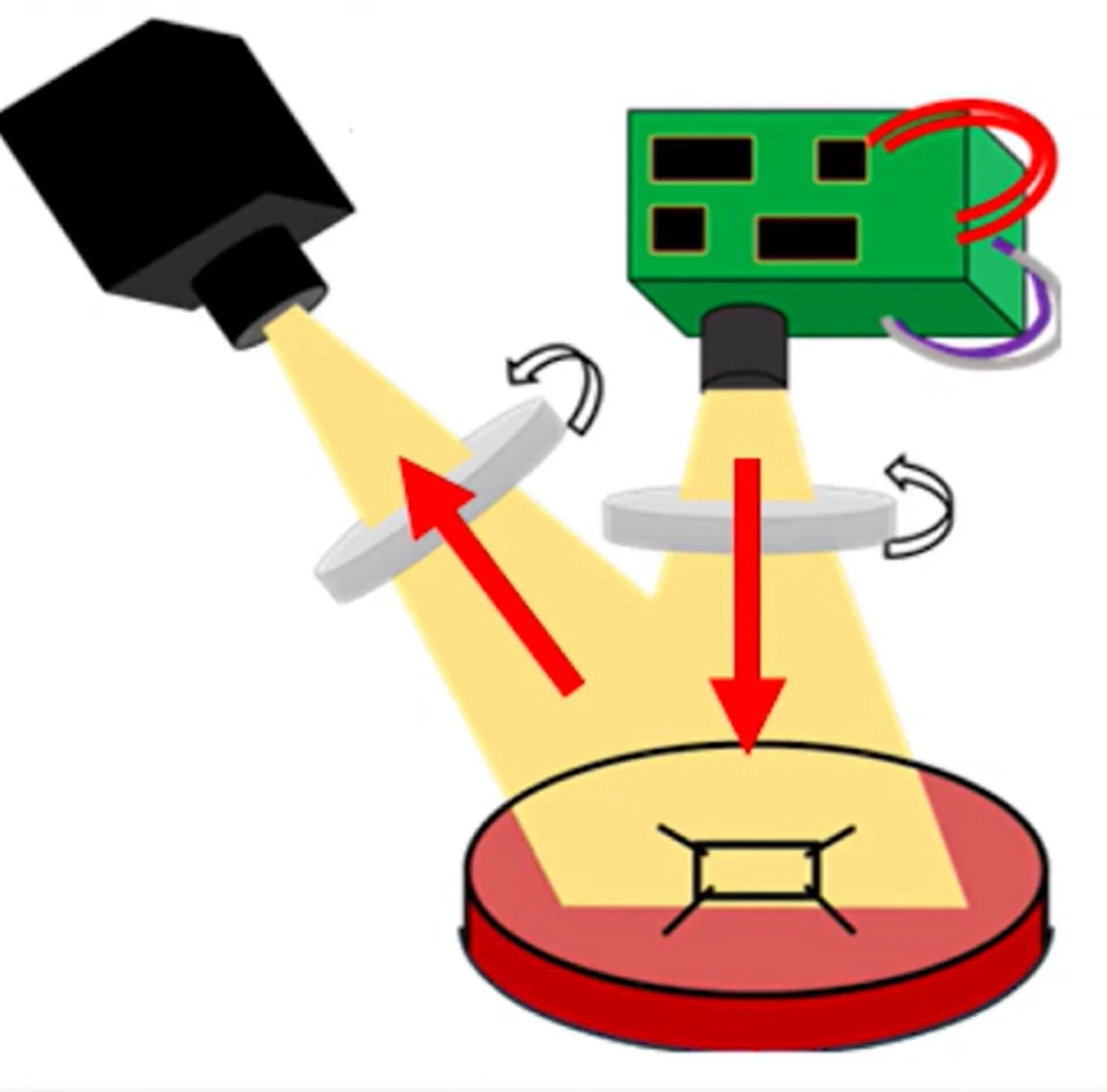
Working alongside CellScale’s mechanical tester, the research team developed a custom polarization-based optical imaging system. The system exploited polarization-dependent light refraction to quantify collagen fiber orientation across the tissue surface.
Key imaging components included:
- Independent force and displacement control in two orthogonal axes
- Submerged testing capability for hydrated soft tissues
- High-resolution force and displacement measurement
- Compatibility with BioRakes and alternative mounting strategies
This integration allowed dynamic imaging of collagen microstructure throughout the loading cycle.
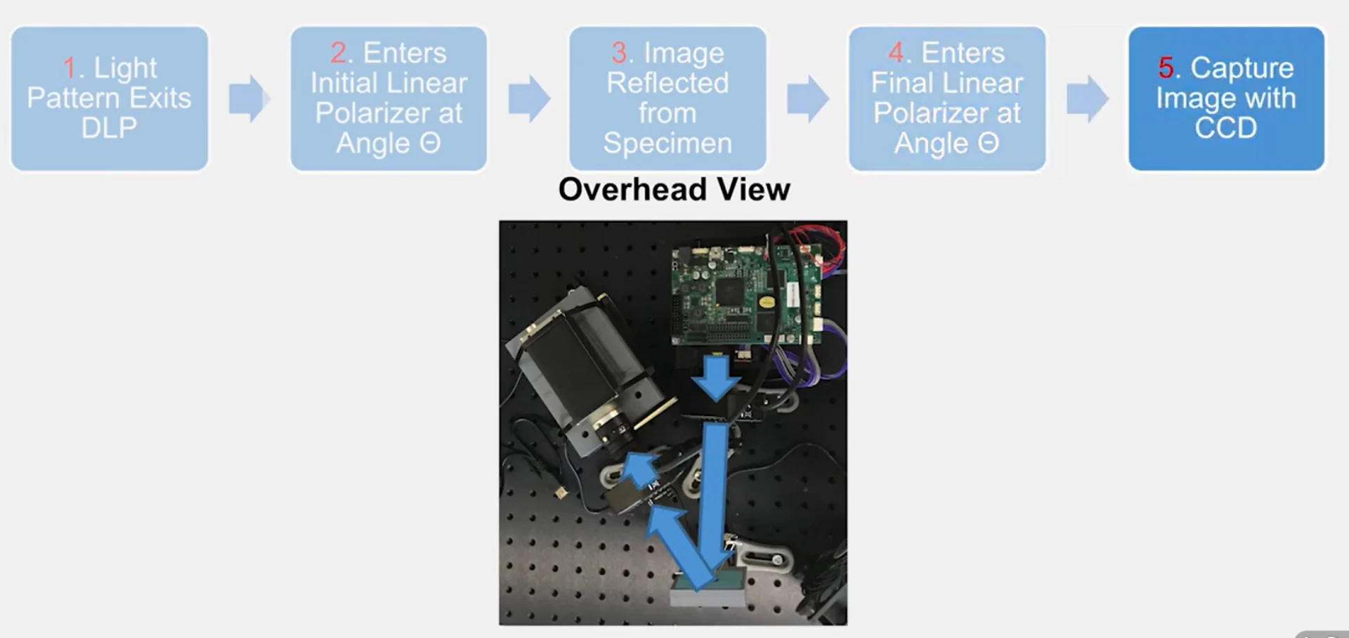
Results and Scientific Impact
Simultaneous Mechanical and Microstructural Data
The integrated system enabled direct correlation between applied mechanical loads and evolving collagen fiber orientation, providing insights not accessible through mechanical testing alone.
Improved Constitutive Model Calibration
Microstructural data supported calibration of advanced hyperelastic models, including anisotropic strain energy functions, improving predictive accuracy in finite element simulations of heart valve behaviour.
Validation of Mounting and Loading Strategies
By comparing BioRake and pulley-based attachment methods, the team assessed how boundary conditions influence local strain distributions and model outcomes.
Key Capabilities Enabled
Heart valve biaxial testing for cardiovascular tissue biomechanics
Real-time collagen fiber orientation measurement
Integrated imaging mechanical testing
Support for advanced constitutive modeling
Customizable experimental configurations
Videos & Gallery


Related Publications
TITLE
Mechanics of Porcine Heart Valves’ Strut Chordae Tendineae Investigated as a Leaflet–Chordae–Papillary Muscle Entity
JOURNAL
Annals of Biomedical Engineering
APPLICATIONS
RESEARCH SUMMARY
Porcine atrioventricular valve strut chordae tendineae were tested using a configuration that retained leaflet, chordae, and papillary muscle attachments during loading. This setup was used to preserve native anatomical interactions during mechanical loading and to better reflect cardiovascular tissue biomechanics.
Mechanical testing was performed primarily under uniaxial loading, with results considered in the context of heart valve biaxial testing approaches used for soft cardiac tissues. Nonlinear and direction-dependent stress–stretch responses were observed, along with regional differences between mitral and tricuspid valves. Mitral strut chordae showed greater thickness and extensibility than tricuspid samples.
Experimental data were fit using an Ogden hyperelastic model to obtain constitutive parameters for computational modeling. Comparisons indicated that retaining leaflet and papillary muscle attachments influenced measured chordal mechanics and altered the resulting material properties used in heart valve simulations.
Citation: Ross, C.J., Laurence, D.W., Hsu, MC. et al. Mechanics of Porcine Heart Valves’ Strut Chordae Tendineae Investigated as a Leaflet–Chordae–Papillary Muscle Entity. Ann Biomed Eng 48, 1463–1474 (2020). https://doi.org/10.1007/s10439-020-02464-6
TITLE
Strain Energy Density as a Gaussian Process and Its Utilization in Stochastic Finite Element Analysis: Application to Planar Soft Tissues
JOURNAL
Computer Methods in Applied Mechanics and Engineering
APPLICATIONS
RESEARCH SUMMARY
Planar soft tissue mechanics were modeled using a data-driven constitutive framework in which strain energy density was represented with a Gaussian process. The formulation was developed to work directly with heart valve biaxial testing data, allowing mechanical response and associated uncertainty to be captured without assuming a predefined material model.
The approach was evaluated using synthetic datasets and experimental biaxial measurements from porcine aortic valve leaflet tissue. Model behaviour was compared with conventional hyperelastic formulations commonly used in valve constitutive modeling, with differences observed in predictive response under multiaxial loading.
The resulting constitutive descriptions were incorporated into finite element simulations, where stochastic methods were used to propagate experimental uncertainty through tissue-level mechanical predictions.
Citation: Ankush Aggarwal, Bjørn Sand Jensen, Sanjay Pant, Chung-Hao Lee. Strain energy density as a Gaussian process and its utilization in stochastic finite element analysis: Application to planar soft tissues. Computer Methods in Applied Mechanics and Engineering. Volume 404, 2023. https://doi.org/10.1016/j.cma.2022.115812
TITLE
A Bayesian constitutive model selection framework for biaxial mechanical testing of planar soft tissues: Application to porcine aortic valves
JOURNAL
Journal of the Mechanical Behavior of Biomedical Materials
APPLICATIONS
RESEARCH SUMMARY
Planar soft tissue mechanics were evaluated using biaxial experimental data collected from porcine aortic valve cusp samples. Mechanical loading was performed under multiple stress ratios, with deformation measured using integrated imaging mechanical testing to capture in-plane tissue response during heart valve biaxial testing.
The resulting datasets were analyzed using principal component analysis and Bayesian probability methods to compare the relative likelihood of several hyperelastic constitutive formulations. Model selection was based on how well each formulation represented variability across samples and loading conditions, rather than on deterministic curve fitting alone.
Across the set of candidate models, the May–Newman formulation showed the highest likelihood for describing the observed biaxial response. The framework provides a structured approach for comparing constitutive descriptions of valve tissue mechanics using probabilistic criteria.
Citation: Ankush Aggarwal, Luke T. Hudson, Devin W. Laurence, Chung-Hao Lee, Sanjay Pant. A Bayesian constitutive model selection framework for biaxial mechanical testing of planar soft tissues: Application to porcine aortic valves. Journal of the Mechanical Behavior of Biomedical Materials. Volume 138, 2023. https://doi.org/10.1016/j.jmbbm.2023.105657
Interested in a Similar Custom Solution?
CellScale supports custom integration of mechanical testing systems with imaging, sensing, and control technologies for advanced biomechanics research.