

Digital Image Correlation
Imaging-Based Mechanical Testing

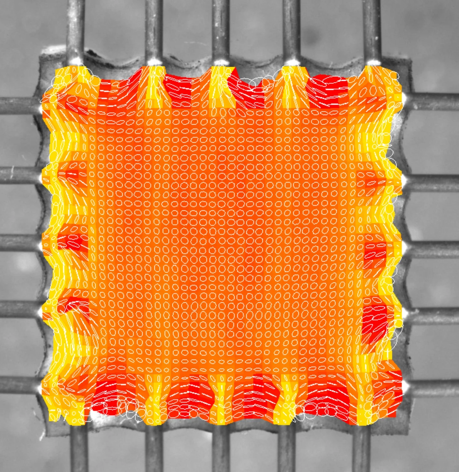
Digital image correlation (DIC) is an optical method used to measure full field strain and deformation during mechanical testing. Imaging-based testing is especially valuable for soft tissues and biomaterials that exhibit nonuniform deformation, anisotropy, or localized strain concentrations that cannot be captured by bulk force and displacement data alone.

What Digital Image Correlation Measures
Digital image correlation tracks surface features or applied speckle patterns to compute deformation across a specimen. This approach enables accurate DIC strain measurement across heterogeneous specimens subjected to complex loading.
DIC Measurement Examples
- Full field strain and displacement
- Local strain gradients and heterogeneity
- Anisotropic deformation behaviour
- Strain concentration and failure initiation
- Time-dependent deformation during loading
- Coupling between force and local strain
- Spatial strain evolution under cyclic loading
DIC provides spatially resolved mechanical information that complements traditional force-based measurements.
Digital Image Correlation in Biomaterials Research
Imaging-based mechanical testing is widely used in:
- Soft tissue biomechanics
Cardiac, vascular, tendon, and ligament tissues often deform nonuniformly under load.
- Engineered tissue characterization
Visualization of strain distribution across scaffolds and tissue constructs, allowing researchers to directly visualize load transfer and strain localization.
- Hydrogel and polymer mechanics
Soft materials frequently exhibit localized deformation that is not reflected in bulk measurements.
- Anisotropy and fibre alignment studies
Full field strain analysis reveals direction dependent behaviour in fibrous materials.
- Model validation and computational biomechanics
DIC data improves calibration of finite element and constitutive models.
Common Sample Types for Imaging-Based Testing
- Soft biological tissues
- Engineered tissue constructs
- Hydrogels and polymer networks
- Membranes and thin films
- Elastomeric materials
- Microtissues and spheroids
- Soft robotics components
- Fibre reinforced biomaterials
How Digital Image Correlation Works
Digital image correlation uses optical imaging synchronized with mechanical loading. This workflow forms the foundation of imaging based mechanical testing, where spatial deformation data are captured alongside force and displacement measurements.
Surface Preparation and Imaging
A natural texture or applied speckle pattern is tracked using high resolution cameras.
Non-Contact Deformation Measurement
This non-invasive approach enables non-contact deformation measurement without mechanically perturbing delicate or hydrated specimens.
Full Field Strain Analysis
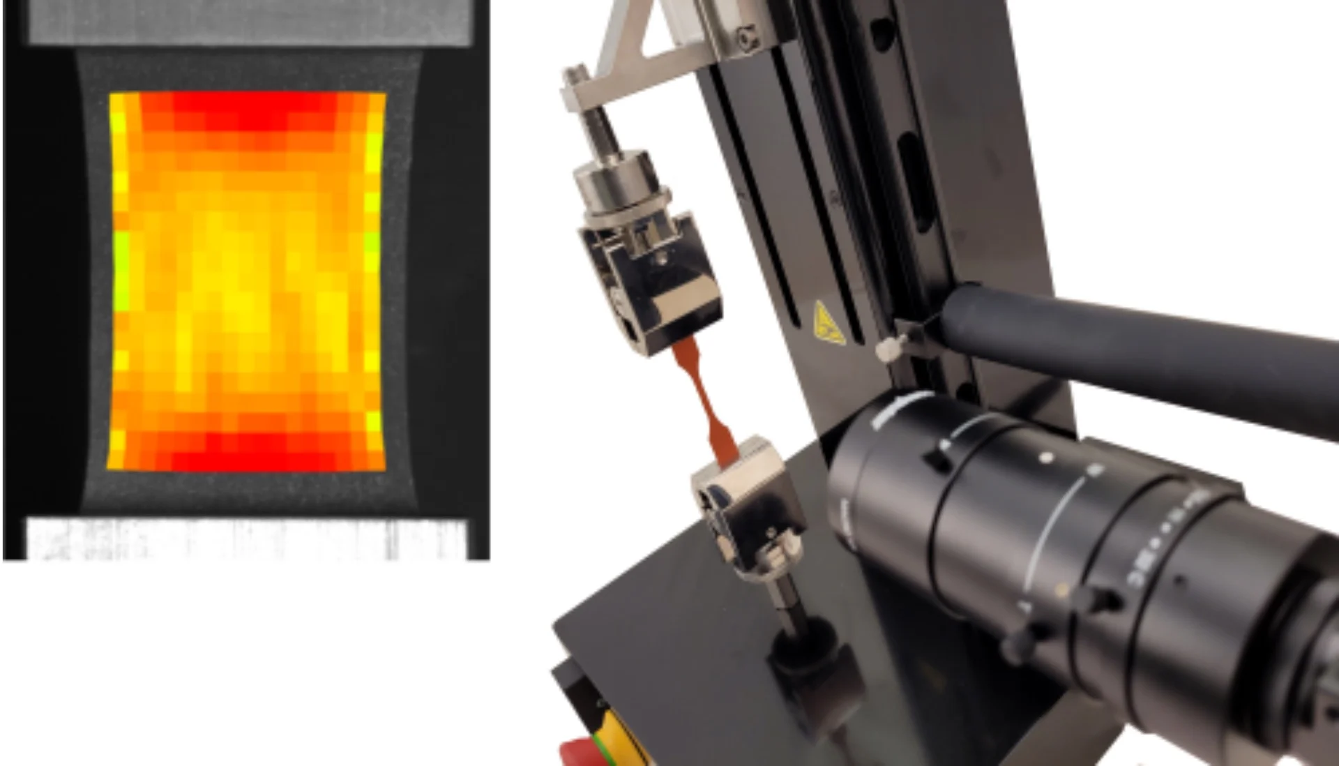
Full field strain analysis generates spatial strain maps across the entire specimen surface, capturing localized deformation and strain gradients.
Time Resolved Imaging
DIC captures strain evolution during static, cyclic or dynamic loading.
Integration with Mechanical Data
Force, displacement, and imaging data are synchronized for comprehensive analysis.
Recommended CellScale Instruments for DIC and Imaging-Based Testing
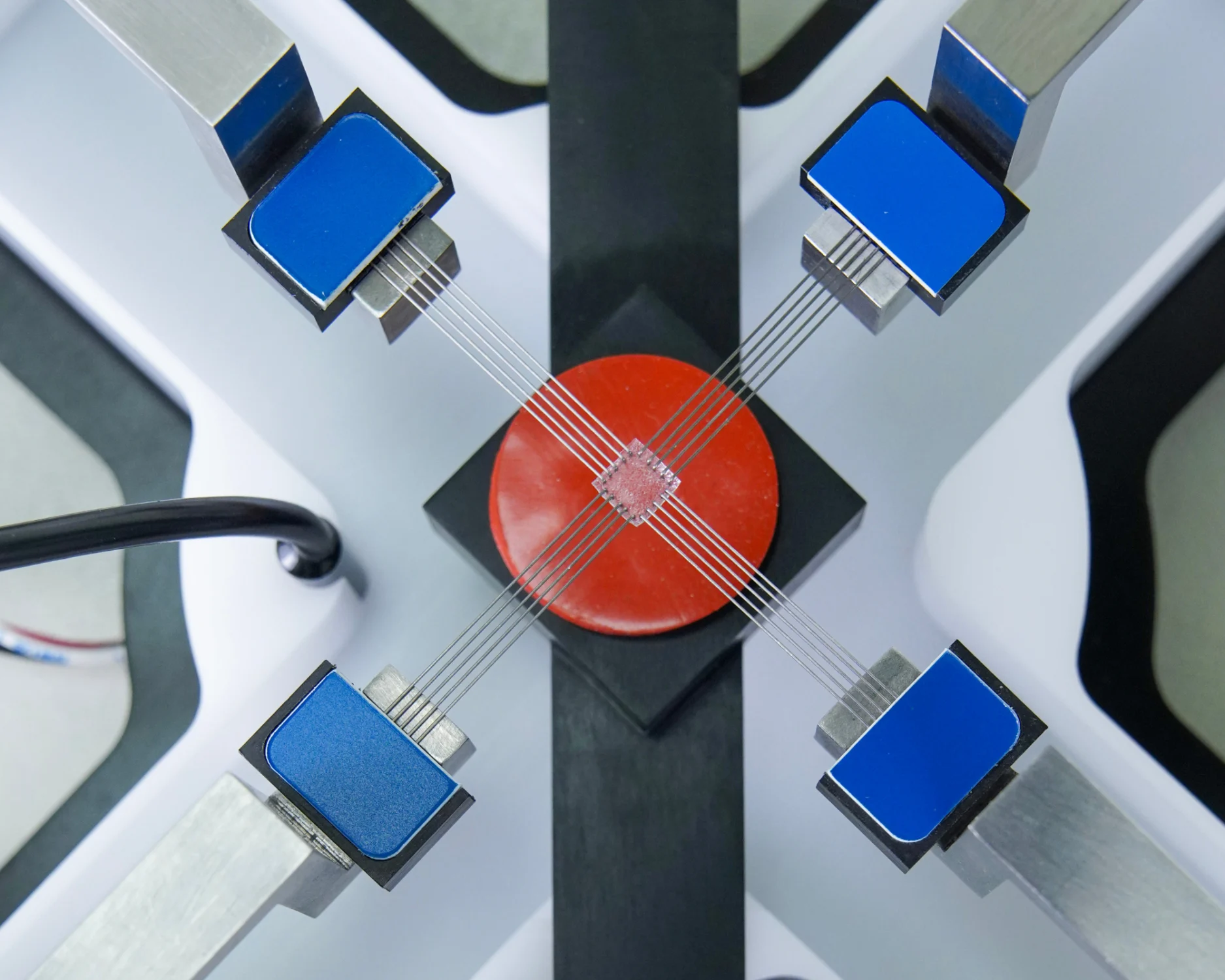
Multiple CellScale instruments integrate mechanical loading with digital image correlation to support imaging-based mechanical testing and full field strain analysis.
BioTester
UniVert
Relevant Research Applications
Digital image correlation and imaging-based testing support research in:
Recent Publications Using Digital Image Correlation
Related Testing Methods
DIC enhances many mechanical tests through full-field strain measurement.
Ready to Perform Imaging-Based Mechanical Testing?
CellScale systems integrate imaging and mechanical loading to deliver precise digital image correlation and full field strain analysis for soft tissues and biomaterials.