High Throughput Micro-Mechanical Testing for Engineered Fibres and Microtissues

A horizontal MicroTester custom configuration enabled micro-scale tensile testing directly in a 96 well plate, supporting high throughput evaluation of engineered fibres, microwires, and microtissues for organ-on-chip applications.
A full side view of the MicroSquisher legacy product
PARTNER INSTITUTION

University of Toronto

Department of Chemical Engineering and Applied Chemistry

Project Background

Engineered fibres, microwires, and microtissues are increasingly used in organ-on-chip systems and advanced biomaterials research. These constructs are typically extremely soft and fragile, often cultured directly within 96 well plates. Standard uniaxial testing systems cannot apply micro-Newton forces without removing constructs from their culture environment or compromising sample integrity.

Researchers in Prof. Milica Radisic’s group at the University of Toronto required a high throughput micro mechanical testing system capable of:

Commercial tensile testers were not designed for this geometry or throughput, prompting the need for a custom engineered micro scale mechanical testing solution.

Headshot of Milica Radisic

Dr. Milica Radisic

Professor & Canada Research Chair, Functional Cardiovascular Tissue Engineering

Figure 2 from the publication that resulted from our high throughput micro mechanical testing work at U of T
Adapted from Y.Zhao, E. Y.Wang, L. H.Davenport, Y.Liao, K.Yeager, G.Vunjak-Novakovic, M.Radisic, B.Zhang, Adv. Healthcare Mater.2019, 8, 1801187. https://doi.org/10.1002/adhm.201801187

The Challenge

Orientation Constraint

Fibres were cultured horizontally within wells. A vertical test configuration would alter geometry, introduce artifacts or damage constructs.

The system needed to preserve the MicroTester’s ability to resolve extremely small forces during tension testing.

96 well plate testing needed to occur with minimal sample disturbance.

Microscopy access was required to monitor fibre deformation and morphology during loading.

Scaffolds, microwires, and microtissues could not be clamped aggressively or manipulated outside their native culture environment.

Custom Solution Developed by CellScale

Horizontal Reorientation of the Force-Sensing Beam

The core micro force beam was rotated into a horizontal configuration, enabling controlled tensile loading while keeping fibres suspended across their native wells.

Direct 96 Well Plate Testing Compatibility

A custom mounting interface allowed the MicroTester to position accurately over each well for rapid, repeatable tension tests.

Custom Micro-Grips and Hooks

Low-mass, compliant hooks were developed to secure microfibres and engineered tissues without altering their geometry or causing damage.

Integrated Imaging Access

The horizontal layout maintained optical access for imaging during testing, supporting deformation tracking and strain quantification.

Hydrated Testing Environment

Testing could be performed directly in media, preserving physiological conditions.

This configuration preserved the MicroTester’s micro-Newton sensitivity while adding new orientation and throughput capabilities tailored to the lab’s workflow.

Results and Scientific Impact

The custom system became a foundational tool for multiple high-impact research programs focused on engineered cardiac tissues, flexible microelectrodes, elastic microwires, and microphysiological systems.

Figure 4 from the publication that resulted from our high throughput micro mechanical testing work at U of T
Adapted from Y.Zhao, E. Y.Wang, L. H.Davenport, Y.Liao, K.Yeager, G.Vunjak-Novakovic, M.Radisic, B.Zhang, Adv. Healthcare Mater.2019, 8, 1801187. https://doi.org/10.1002/adhm.201801187

Enabled high throughput micro tensile testing

Dozens of samples could be tested rapidly without culture disruption.

Supported development of heart-on-a-chip platforms

Mechanical characterization of microwires and tissue fibres informed device design and biological performance.

Advanced studies in cardiac fibrosis and tissue remodeling

Micro tensile testing quantified stiffness changes and maturation over time.

Facilitated innovation in printed elastomeric and composite materials

Mechanical evaluation guided optimization of flexible polymer electrodes and multi-material structures.

Across multiple peer-reviewed publications, the custom MicroTester configuration supported studies spanning engineered cardiac tissues, microwires, and microphysiological systems.

Key Capabilities Enabled

Horizontal micro tension testing with rapid multi-sample workflows

Micro-Newton force sensitivity with custom micro-grip interfaces

Hydrated, imaging-compatible test environment

Precise displacement control for micro-scale mechanics

Video & Gallery

Engineered Fiber Micro-Mechanical Testing

Micro-scale tensile testing in a 96 well plate using a custom horizontal MicroTester configuration, with compliant hooks and optical access for deformation tracking and strain quantification.

Related Publications

TITLE

A Multimaterial Microphysiological Platform Enabled by Rapid Casting of Elastic Microwires

JOURNAL

Advanced Healthcare Materials

APPLICATIONS

RESEARCH SUMMARY

A 96 well plate testing platform was developed for use with engineered human cardiac tissues. The system supports high throughput micro mechanical testing by combining force sensing and electrical stimulation within a scalable microwell format.

Soft elastic POMaC microwires were used as both tissue anchors and force sensors, allowing repeated, non-invasive measurements during culture. Mechanical characterization included passive micro tensile testing, active force generation, and contraction–relaxation behavior collected over extended time periods.

Carbon electrodes were integrated into the platform to enable chronic electrical pacing during tissue maturation. Functional responses were evaluated under controlled conditions, including pharmacological perturbation, to assess consistency and sensitivity across samples.

Citation: Citation: Y. Zhao, E. Y. Wang, L. H. Davenport, Y. Liao, K. Yeager, G. Vunjak-Novakovic, M. Radisic, B. Zhang, Adv. Healthcare Mater. 2019, 8, 1801187. https://doi.org/10.1002/adhm.201801187

TITLE

A Platform for Generation of Chamber-Specific Cardiac Tissues and Disease Modeling

JOURNAL

Cell

APPLICATIONS

RESEARCH SUMMARY

This study presents the Biowire II platform, a micro scale mechanical testing cardiac tissue system that enables long-term culture and non-invasive, online measurement of micro tensile testing, active force, contractile dynamics, and calcium transients under electrical pacing. Using directed differentiation plus chronic electrical conditioning, the authors generated electrophysiologically distinct atrial and ventricular tissues with chamber-specific gene expression and drug responses, and engineered heteropolar atrio-ventricular tissues with spatially confined responses to serotonin and ranolazine. The platform also supported months-long conditioning (up to ~8 months) to model polygenic cardiac disease (left ventricular hypertrophy) from patient-derived iPSCs.

Citation: Zhao, Y., Rafatian, N., Feric, N. T., Cox, B. J., Aschar-Sobbi, R., Wang, E. Y., … Radisic, M. (2019). A platform for generation of chamber specific cardiac tissues and disease modelling. Cell, 176(4), 917–930.e17. https://doi.org/10.1016/j.cell.2018.11.042

TITLE

Heart-on-a-Chip Model of Epicardial–Myocardial Interaction in Ischemia Reperfusion Injury

JOURNAL

Advanced Healthcare Materials

APPLICATIONS

RESEARCH SUMMARY

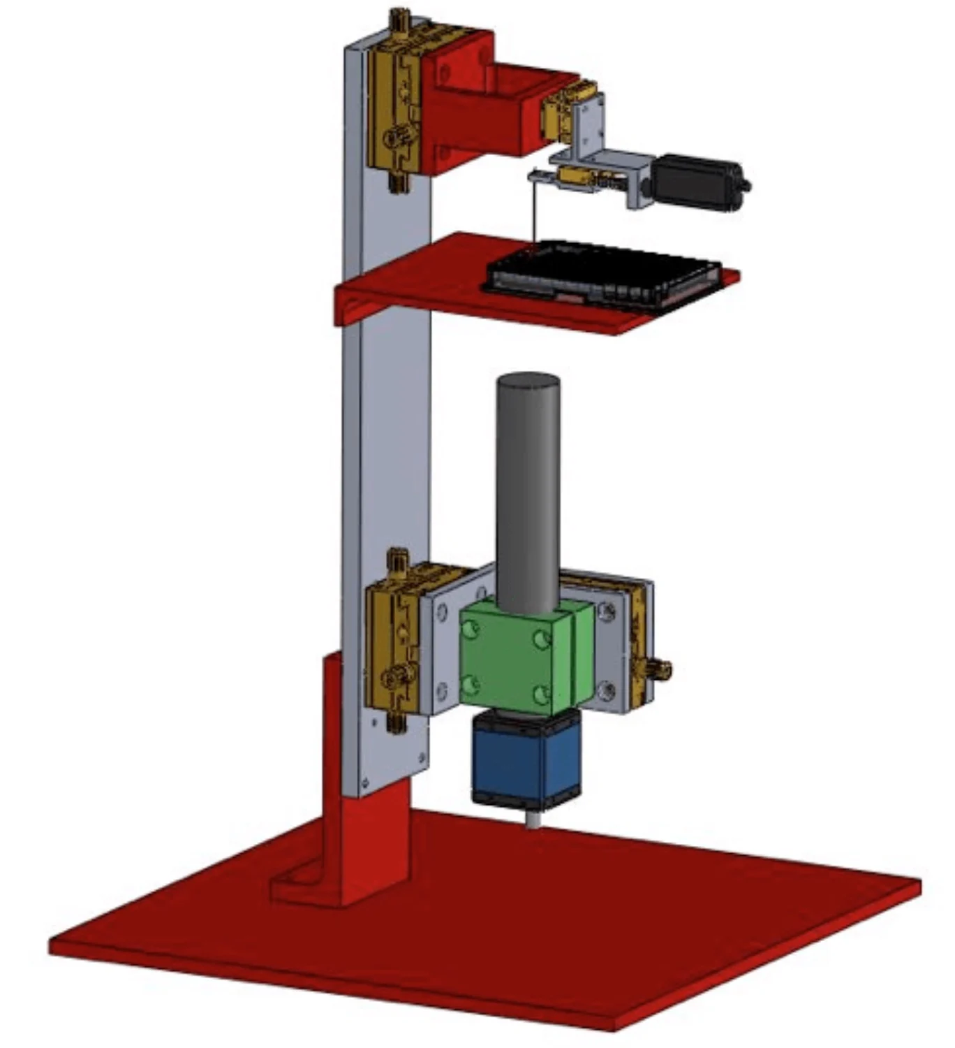
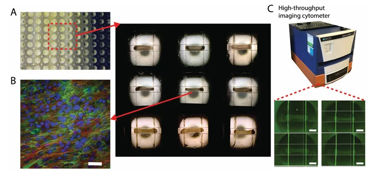
A heart-on-a-chip platform was developed to study interactions between epicardial and myocardial tissue layers under controlled conditions. The system combined engineered epicardial tissue with a myocardial core, enabling measurements of tissue structure and function using micro scale mechanical testing approaches compatible with high throughput micro mechanical testing workflows.

Human pluripotent stem cell–derived cardiomyocytes, fibroblasts, and epicardial cells were introduced using a two-step seeding process. Over time, epicardial cells migrated into the myocardial layer, forming bilayer tissues with maintained organization and evolving functional behavior during culture.

Tissues were subjected to an ischemia–reperfusion injury protocol to examine differential responses. Samples containing an epicardial layer showed reduced cell death and distinct functional recovery patterns compared to myocardial-only constructs. Quantitative imaging, force measurements, and immunostaining were used to track epicardial cell behavior and mechanical response during and after injury.

This platform supports the study of epicardial–myocardial interactions across development and injury models, with mechanical measurements integrated alongside biological readouts.

The resulting datasets were analyzed using principal component analysis and Bayesian probability methods to compare the relative likelihood of several hyperelastic constitutive formulations. Model selection was based on how well each formulation represented variability across samples and loading conditions, rather than on deterministic curve fitting alone.

Across the set of candidate models, the May–Newman formulation showed the highest likelihood for describing the observed biaxial response. The framework provides a structured approach for comparing constitutive descriptions of valve tissue mechanics using probabilistic criteria.

Citation: D. Bannerman, S. Pascual-Gil, Q. Wu, I. Fernandes, Y. Zhao, K. T. Wagner, S. Okhovatian, S. Landau, N. Rafatian, D. F. Bodenstein, Y. Wang, T. R. Nash, G. Vunjak-Novakovic, G. Keller, S. Epelman, M. Radisic, Heart-on-a-Chip Model of Epicardial–Myocardial Interaction in Ischemia Reperfusion Injury. Adv. Healthcare Mater. 2024, 13, 2302642. https://doi.org/10.1002/adhm.202302642

Interested in a Similar Custom Solution?

CellScale engineers collaborate directly with research teams to design custom testing configurations like the high throughput micro mechanical testing system for U of T, that meet specialized scientific needs.

Contact Sales

Product of Interest: