


Arteries do not sit loosely in the body. They are held under pressure, but they are also stretched along their length. That second part is easy to overlook, especially when arterial tissue is removed, cut open, and tested as a flat specimen.
In a study published in the Journal of the Mechanical Behavior of Biomedical Materials, researchers used the CellScale BioTester to measure the biaxial mechanics of human common carotid arteries. They then combined those measurements with each artery’s axial pre-stretch to simulate how the intact vessel might respond to inflation and extension.
The work is an interesting example of how carotid artery biaxial testing can feed into a more physiological model, even when the experiment itself begins with a small, flattened piece of tissue.
Why Carotid Artery Biaxial Testing Needs Two Loading Directions
The common carotid artery carries blood toward the brain and facial tissues. Its wall contains elastin, collagen, and other structural components arranged in several directions. As a result, pulling the artery circumferentially can affect its axial response, and axial stretching can change how it behaves under pressure.
A conventional tensile test captures only one direction at a time. Biaxial testing applies controlled loading along two axes, making it possible to measure the directional coupling that appears in arterial tissue.
That matters when the data will eventually be used in a constitutive model. A model based on a single tensile direction may fit that one test well but still miss what happens when pressure and axial stretch act together.
The authors were also working with human donor tissue, where sample availability is limited. Planar arterial tissue biaxial testing only requires a small section of the vessel. Tubular inflation testing, by comparison, needs an intact segment that can be sealed and pressurized without leaking. In practice, that is not always available.
Preparing Human Carotid Arteries and Measuring Axial Pre-Stretch
Left and right common carotid arteries were collected from 10 human donors. Nine donors were between 78 and 98 years old. One donor was 19 years old.
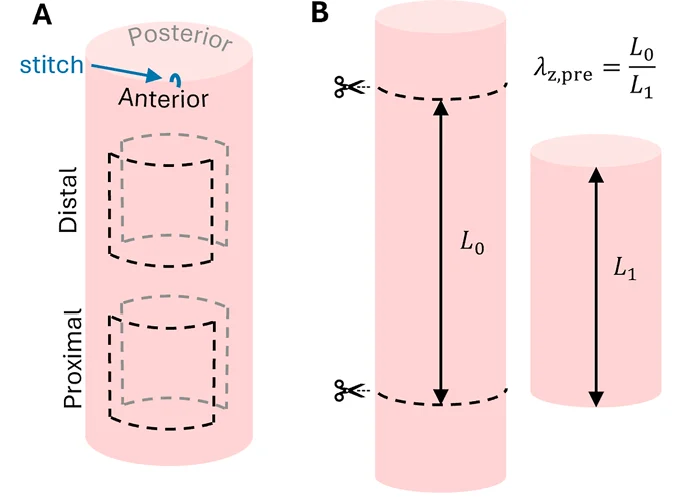
Before each artery was removed, the researchers marked two locations along its length. They measured the distance between those marks before and after excision. The ratio between those measurements was used to estimate the artery’s in situ axial pre-stretch.
The team also kept track of anterior, posterior, proximal, and distal orientations. Square samples of approximately 10 by 10 mm were then cut from the vessel wall. Areas with visible calcification, fractures, or other obvious damage were avoided.
This orientation work is more than record keeping. If one side of the artery were consistently stiffer than another, or if the proximal region behaved differently from the distal region, those differences could affect how confidently the tissue data are generalized.
How the BioTester Was Used for Carotid Artery Biaxial Testing
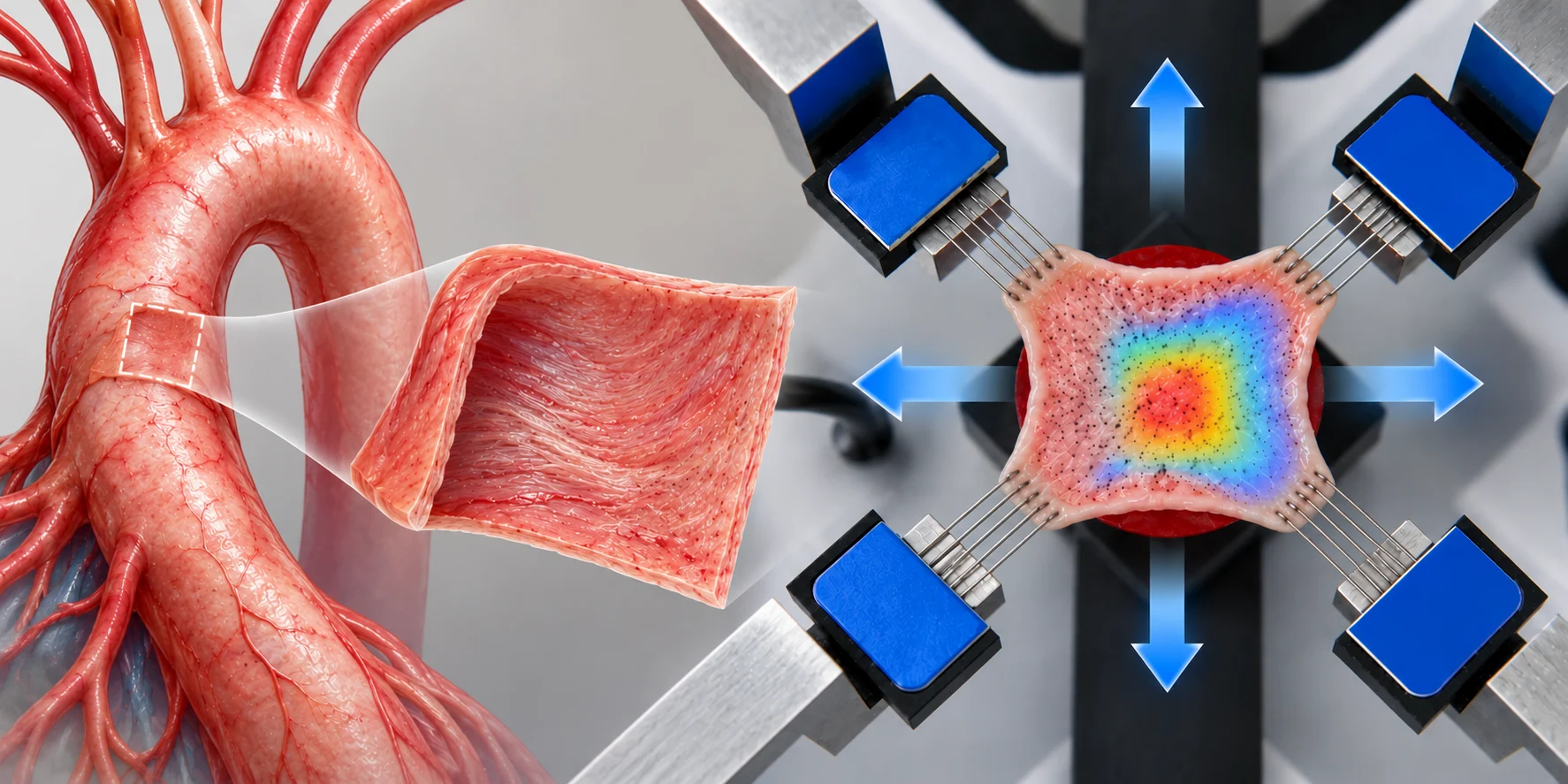
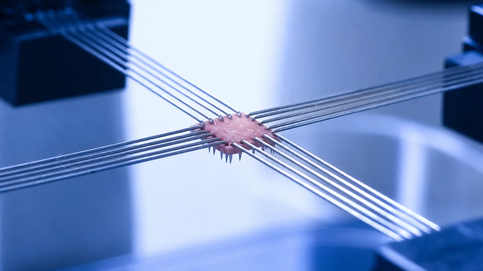
The researchers mounted each arterial sample in a BioTester 5000 using CellScale BioRakes. Five small hooks engaged each edge of the tissue, allowing the specimen to be pulled in the circumferential and axial directions.
The intimal surface faced upward and was marked with graphite speckles. Samples remained submerged in phosphate-buffered saline maintained at 37°C throughout testing, a setup consistent with hydrated testing of soft biological tissues.
The testing protocol began with six force-controlled equibiaxial preconditioning cycles. The researchers then switched to stretch-controlled loading and applied several circumferential-to-axial stretch ratios. These ranged from equal loading in both directions to strongly biased conditions such as 1:0.25 and 0.25:1.
A 20 mN preload was applied at the beginning of each loading cycle. Forces and images were recorded at 10 Hz.
Local tissue deformation was calculated in our LabJoy software using a 7 by 7 tracking grid. The analysis focused on the central 30% of the specimen to reduce edge and mounting effects. This image-based strain measurement is closely related to the workflows described in digital image correlation, although the authors described their method specifically as local deformation tracking.
One practical detail surfaced during testing. In six samples, the mounting arms contacted the vessel bath and introduced force measurement artefacts. Those samples were excluded, leaving 29 specimens in the final analysis. The paper is unusually open about this sort of problem, which is useful when considering how a similar protocol might be set up.
From Planar Biaxial Data to an Inflating Artery
A flattened artery is not mechanically identical to a closed, pressurized artery.
Cutting the vessel open releases residual stress. Flattening it then stretches some parts of the wall and compresses others. The authors addressed this by fitting a thick-walled Holzapfel-Gasser-Ogden model to the carotid artery biaxial testing data.
The model included the stress-free opened configuration, the flattened testing state, the closed tubular state, and the loaded artery. It was then used to simulate two conditions:
1. Inflation from 0 to 150 mmHg at the measured axial pre-stretch
2. Axial extension from a stretch of 1.0 to 1.2 at a fixed pressure of 100 mmHg
This is where the experimental work moves beyond a set of stress-stretch curves. The BioTester measurements supplied material information across several loading ratios. The model then used that information to estimate diameter, wall thickness, stress, stiffness, reduced axial force, and strain energy under pressure and extension.
A related experimental-to-computational workflow appears in earlier work on mitral valve biomechanics and fluid-structure interaction simulation, where measured biaxial tissue behaviour was carried into a physiological model.
What Did the Authors Measure for Axial Pre-Stretch?
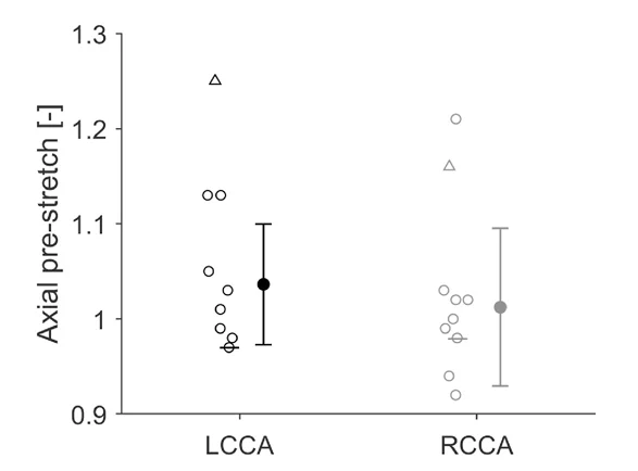
The young donor had left and right carotid axial pre-stretches of 1.25 and 1.16. The older donor arteries were much closer to an unstretched ratio, averaging about 1.03 on the left and 1.01 on the right.
Some older specimens had values below 1.0. In other words, those arteries lengthened rather than shortened when removed. The authors suggest that age-related tortuosity or differences in neck position during dissection may have contributed.
The comparison is visually striking, but it needs some restraint. There was only one young donor, and that donor also experienced a different storage history. The study gives a useful view of how the young samples behaved, though it does not provide a balanced young-versus-aged cohort.
What the Simulated Inflation and Extension Experiments Showed
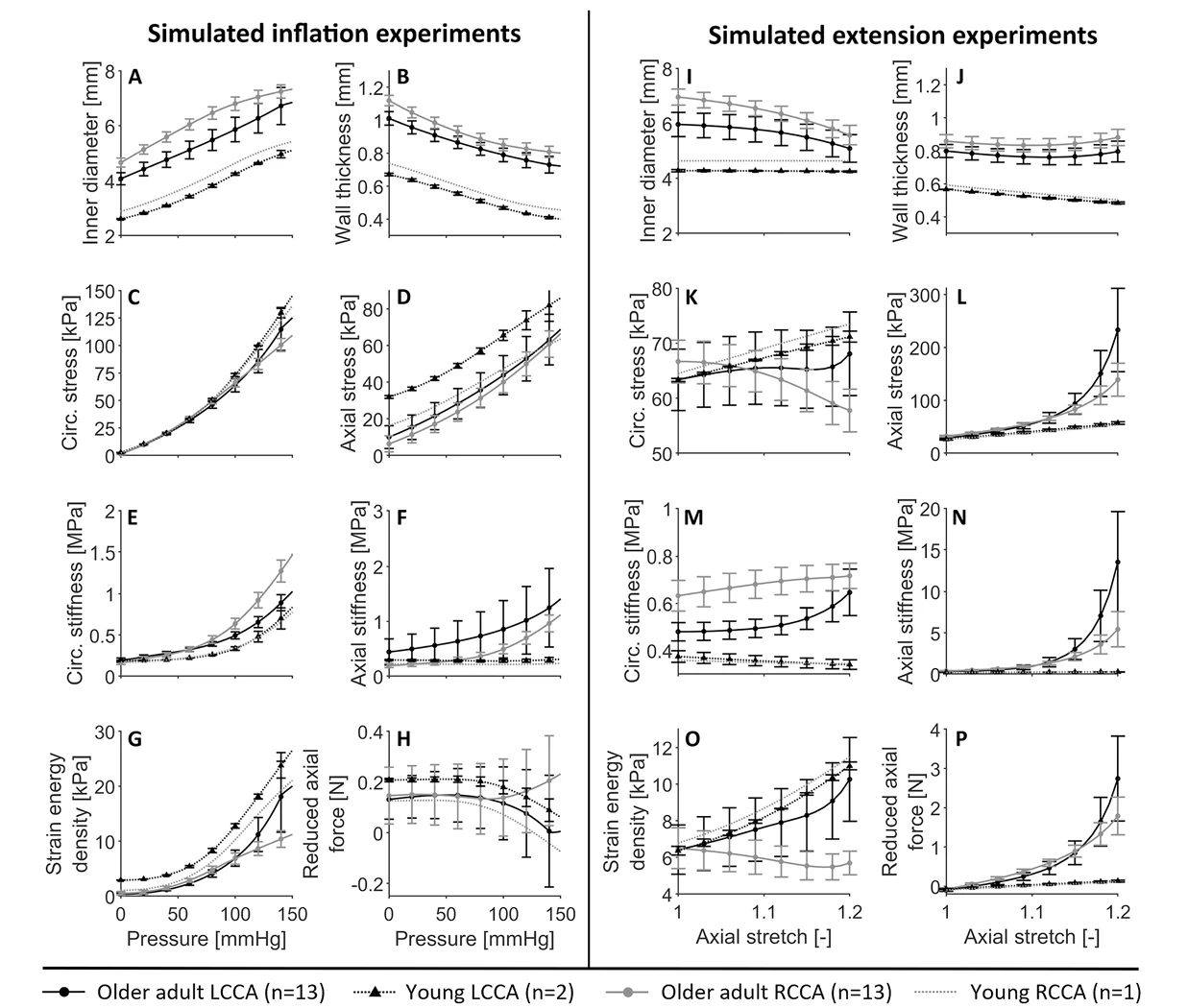
During simulated inflation, the inner diameter increased and the wall became thinner. Circumferential stress rose almost linearly with pressure, while circumferential stiffness increased more sharply at higher pressures.
Axial stress and axial stiffness also increased during inflation, even though the main applied load was pressure. This is one of the more useful outcomes of the study. Axial and circumferential mechanics were not acting independently.
The reduced axial force remained relatively steady in the older donor arteries during inflation. That is what would generally be expected when an artery is held near its physiological axial pre-stretch.
The simulated extension tests told a different part of the story. At 100 mmHg, increasing axial stretch caused axial stress, axial stiffness, and reduced axial force to climb rapidly. Circumferential behaviour also shifted, though the patterns were not identical between left and right arteries.
The young donor samples generally had smaller diameters and thinner walls. Their axial stress and stiffness during extension were also lower than those of the older donor samples. Again, it appears consistent with age-related arterial stiffening, but the sample numbers do not support a broad age comparison on their own.
Were the Left and Right Carotid Arteries Mechanically Different?
For the most part, differences between locations were limited.
The right carotid arteries had larger loaded diameters at lower pressures. They also appeared circumferentially stiffer at 100 and 140 mmHg, while axial stiffness was higher on the left at those pressures.
The anterior side was circumferentially stiffer than the posterior side at 60 mmHg, but that difference did not remain significant at higher pressures. Proximal and distal regions were broadly similar.
That relative consistency may be useful when human tissue is scarce. It suggests that one small arterial sample may still provide data that are reasonably representative of the surrounding vessel, at least within the limits of this donor group.
Researchers interested in similar questions across other vascular beds may also want to read CellScale’s earlier overview of vascular biomechanics research using the BioTester. That work includes human arterial ageing, flattening effects, anisotropy, and constitutive modelling in femoropopliteal arteries.
Interpreting Carotid Artery Biaxial Testing Data Carefully
The study has several limitations worth keeping close to the results.
Donor health histories were incomplete, and some donors had conditions including hypertension, thrombosis, or atherosclerosis. Visibly affected areas were avoided, but less obvious tissue changes may still have influenced the mechanics.
All arteries were frozen before testing. Frozen storage can affect wall thickness, extracellular matrix structure, and stiffness. The youngest donor was stored under different conditions and for a much longer period before the arteries were collected.
The axial pre-stretch measurements were also made in cadavers at very low residual pressure. Whether those measurements exactly match the living, pressurized state is uncertain.
There was also a preload issue. Because the 20 mN preload was reapplied before every cycle, the starting stretch varied slightly across the protocol. The authors corrected this during analysis, though they note that future experiments should apply preload once at the beginning.
These details do not undo the carotid artery biaxial testing results. They do affect how far the age comparison, physiological interpretation, and model outputs should be extended.
Where This Approach May Be Useful
The paper is centred on native human carotid arteries, but the workflow also connects to vascular tissue engineering and mechanics.
Researchers developing vascular grafts, patches, decellularized vessels, or tissue-engineered blood vessels often need to compare their constructs against native tissue. Circumferential stiffness alone is rarely enough. Axial behaviour, anisotropy, pressure response, and the interaction between loading directions can all affect how a graft behaves after implantation.
The same BioTester workflow has also been used for other collagen-rich cardiovascular tissues. One recent example examined porcine pericardium biaxial testing for heart valve design under hydrated, physiological-temperature conditions.
Citation
About the BioTester
The CellScale BioTester is used for planar biaxial testing of soft tissues and biomaterials.
Researchers mount specimens between four independently controlled actuators using BioRakes, clamps, hooks, or other attachments. Force is measured in two perpendicular directions while the sample surface is recorded for local strain analysis. Testing can be performed in a fluid bath, including warmed physiological solution.
In this study, the BioTester was used to generate multi-ratio force and deformation data from small pieces of human carotid artery. Those measurements were then fitted to a constitutive model and translated into simulated pressure and extension behaviour.
That is one reason carotid artery biaxial testing is useful beyond the test itself. The measured mechanics can become inputs for models of the closed, loaded vessel, where pressure and axial stretch are acting at the same time.