Clinical Heart Valve Material Quantification for Pediatric Cardiac Surgery
Boston Children’s Hospital
Department of Cardiac Surgery with Dr. Peter Hammer
Project Background
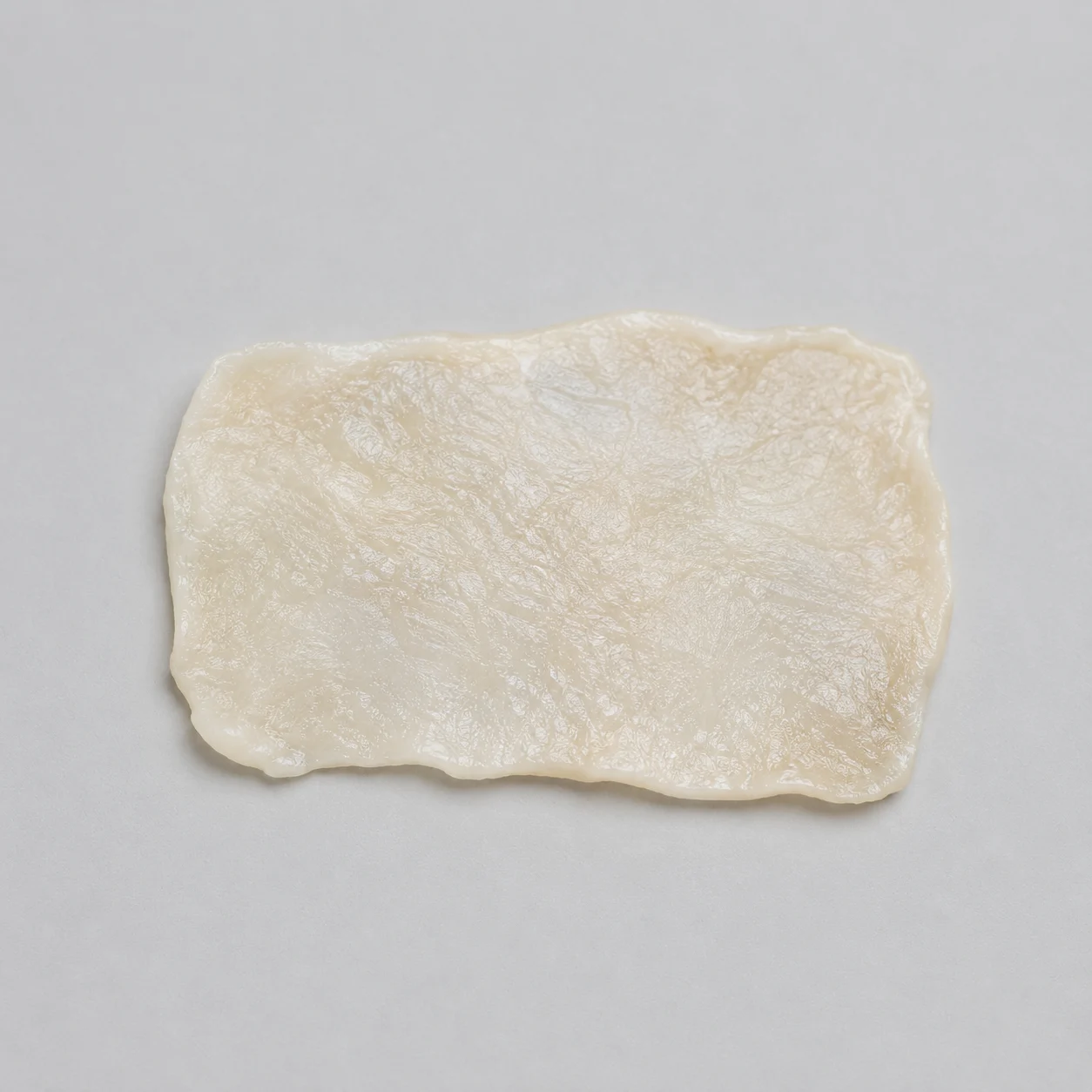
In pediatric heart valve biomechanics, repair differs from adult valve replacement in both approach and constraints. In many cases, surgeons reconstruct valves using the patient’s own tissue, including pericardium, rather than implanting a manufactured replacement. This allows for growth but also introduces variability in material behaviour.
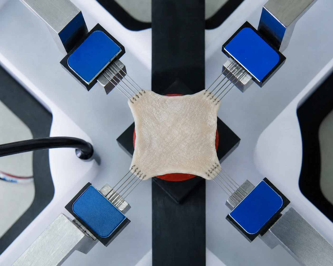
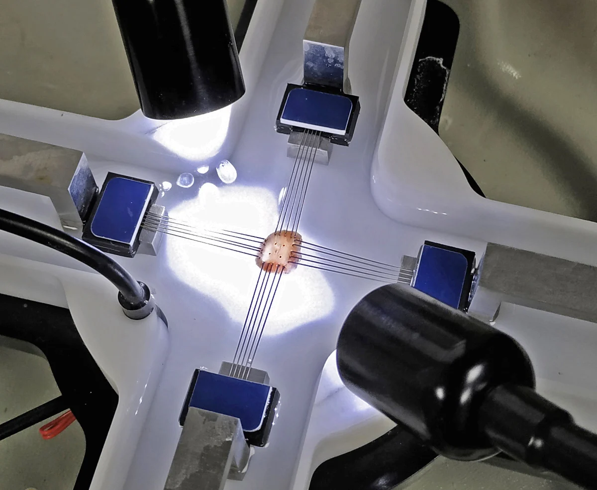
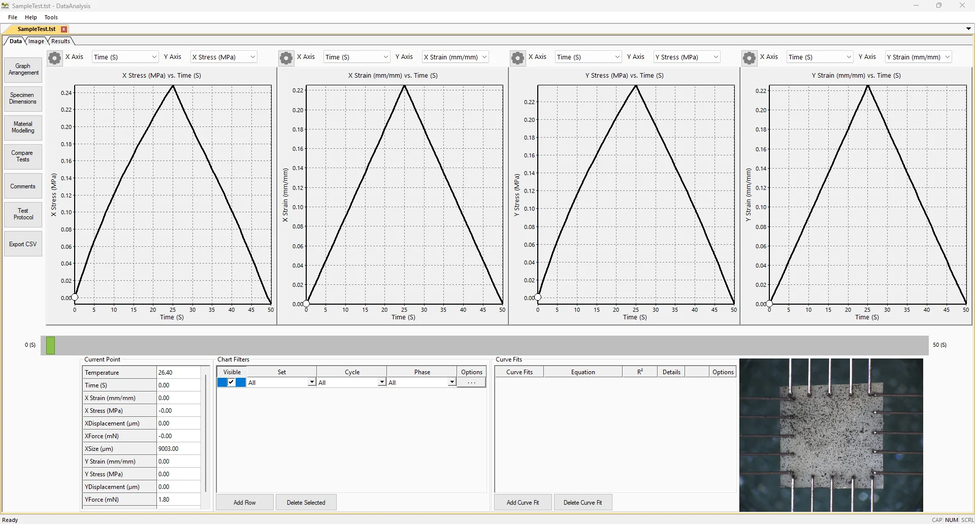
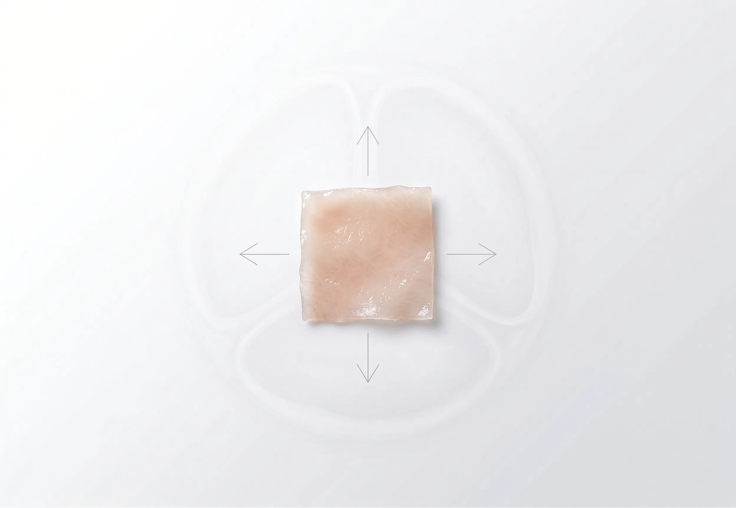
Materials used for valve repair do not always exhibit the same mechanical response as native valve tissue. Differences in stiffness, extensibility, and directional behaviour can affect valve motion and long-term durability after surgery.
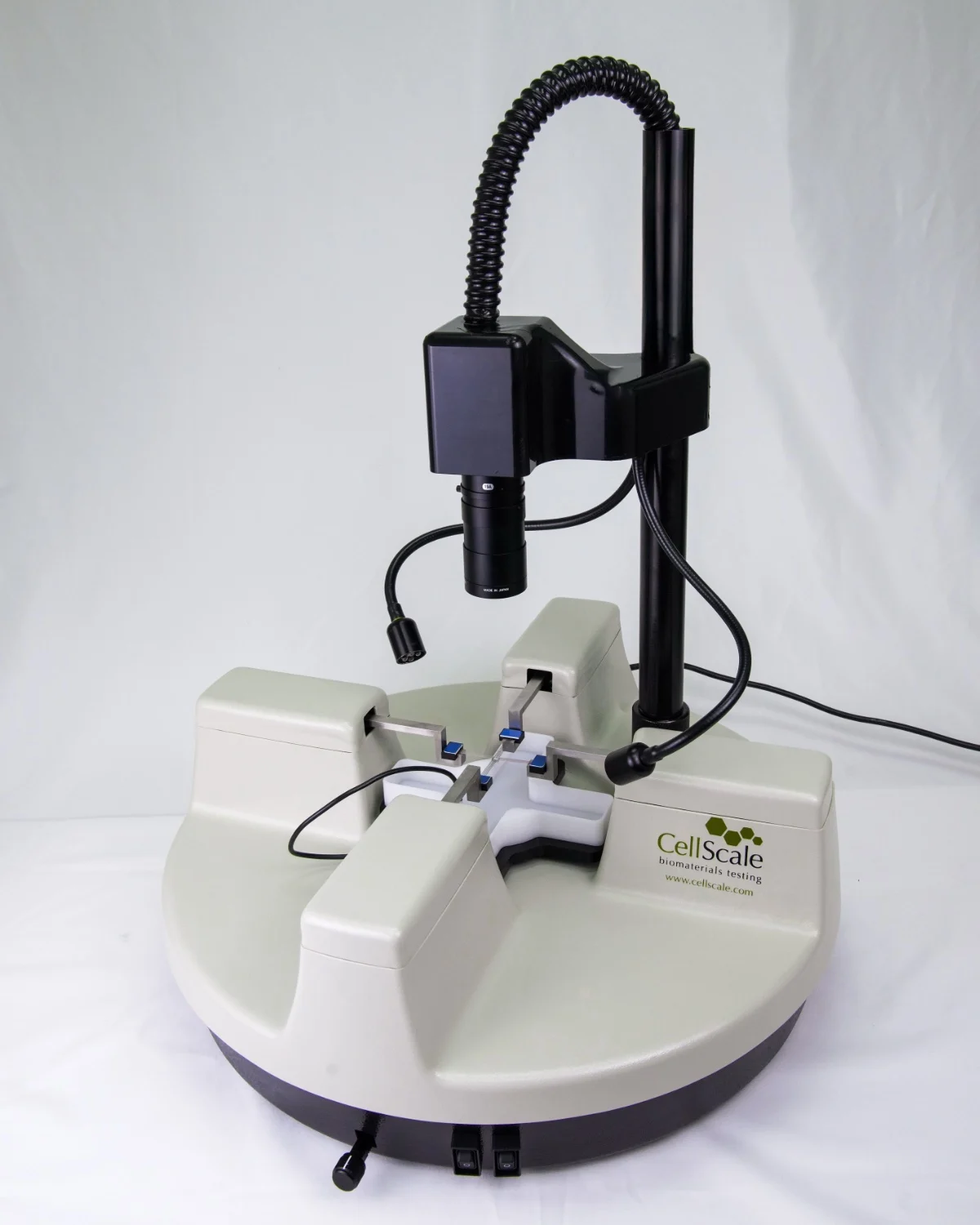
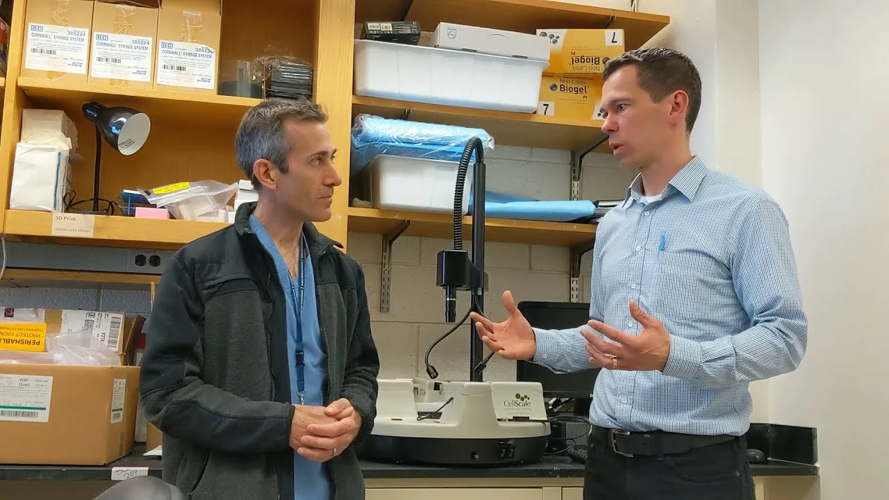
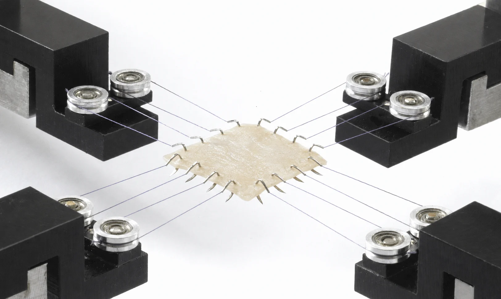
At Boston Children’s Hospital, Dr. Peter Hammer works with cardiac surgeons to incorporate mechanical measurements into the evaluation of valve and repair tissues. Heart valve material quantification data are used to compare native and repair materials and to support surgical decisions in pediatric cardiology.
Dr. Peter Hammer
PhD Co-Directors, CardioEngineering Lab; Scientist, Congenital Heart Valve Program Instructor of Surgery, Harvard Medical School
The Challenge
Variability in Repair Materials
Pericardial tissues harvested during surgery can vary significantly depending on:
- Anatomical harvest location
- Chemical fixation methods and duration
- Orientation and handling during preparation
Surgeons traditionally rely on experience and qualitative assessment to account for these differences.
Translating Engineering Data to the Operating Room
Biomechanical testing methods are commonly used in engineering research, but applying the resulting data during surgical procedures requires simplification and clear interpretation.
Mechanical Testing Approach
Biaxial Mechanical Testing of Repair Tissues
Discarded pericardial and valve tissues from surgical procedures were collected and tested using the CellScale BioTester biaxial mechanical tester. This approach enabled characterization of tissue behaviour under physiologically relevant loading conditions.
Biaxial mechanical testing allowed the team to:
- Properly measure heart valve material quantification
- Quantify tissue extensibility and stiffness
- Compare native valve tissue to treated pericardial patches
- Evaluate the effects of fixation time and tissue orientation
Heart Valve Material Quantification Data for Surgical Guidance
Rather than focusing on material modeling alone, test results were organized into comparative datasets that surgeons could use to understand how different preparation methods influence tissue mechanics.
This integration allowed dynamic imaging of collagen microstructure throughout the loading cycle.
Results and Clinical Impact
Objective Comparison of Repair Materials
Mechanical testing showed measurable differences between native valve tissue and pericardial patches prepared using different processing protocols. Variations in stiffness and extensibility were observed across samples, providing a mechanical basis for differences seen during valve repair procedures.
Improved Surgical Decision-Making
Numerical measurements of tissue stiffness and extensibility were introduced alongside existing surgical assessment methods. These values were used to compare available repair materials and to improve surgical decisions in pediatric cardiology related to patch sizing, shaping, and fixation during heart valve procedures.
Rapid Translation to Clinical Practice
Because testing focused on clinically relevant questions rather than abstract material models, results were disseminated through surgeon-focused publications and direct collaboration with surgical teams. A great example of translational biomechanics at work.
Key Capabilities Enabled
Heart valve material quantification
Direct comparison of treatment and fixation protocols
Translation of mechanical data into surgical guidance
Support for pediatric cardiac surgery research
Related Publication
TITLE
Preclinical Validation of a Patient-Specific Patch-Planning Workflow for Congenital Cardiovascular Reconstruction
JOURNAL
Annals of Biomedical Engineering
APPLICATIONS
RESEARCH SUMMARY
Patient-specific surgical patch planning was examined in the context of congenital cardiovascular reconstruction. Finite-element and analytical models were applied to relate patch geometry to post-repair vessel diameter in simulated branch pulmonary artery stenosis.
Central to the workflow is heart valve material quantification, where pediatric heart valve biomechanics are represented using experimentally derived mechanical properties. Mechanical inputs were obtained from biaxial mechanical testing and incorporated into virtual 3D vessel models that account for pressure-dependent expansion and suture interaction.
Neonatal and child-scale silicone artery models were fabricated to evaluate the approach using different patch materials, including silicone, pericardium, and porcine tissue. Predicted patch dimensions were compared with reconstructed geometries to assess agreement with target vessel size. The workflow provides a translational biomechanics framework for linking mechanical testing data with surgical decisions in pediatric cardiology.
Citation: Kizilski, S.B., Recco, D.P., Davee, J.M. et al. Preclinical Validation of a Patient-Specific Patch-Planning Workflow for Congenital Cardiovascular Reconstruction. Ann Biomed Eng 53, 3505–3522 (2025). https://doi.org/10.1007/s10439-025-03870-4
Interested in Translational Biomechanics?
CellScale systems support many quantitative mechanical testing workflows designed for translational and clinically focused research.