
In our In the Lab blog series, we catch up with researchers using CellScale systems.
In this installment of the blog, Caleb Horst chats with Professor Cheryl Seguin from the University of Western Ontario, Canada. Cheryl is completing research on annulus cells using the MechanoCulture B1.
Caleb Horst: To start with could you tell me a little bit more about the big picture and what your research with annulus cells is all about?
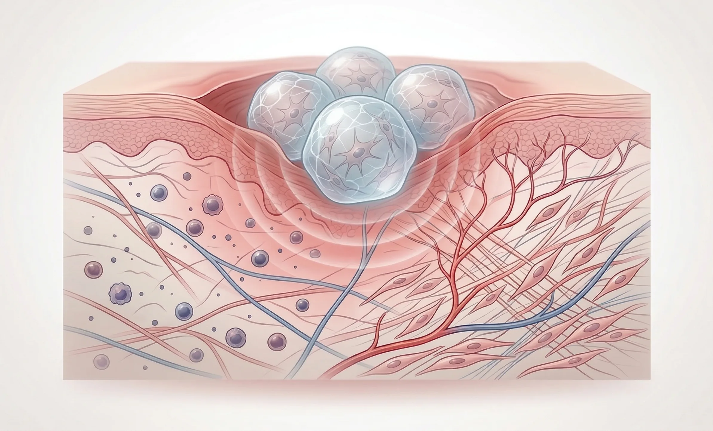
Cheryl Seguin: My lab is focused on understanding the intervertebral discs. We use mouse models to try to understand how the tissue develops, what is important to keep a healthy tissue, and then also how these cells are responding to the complex mechanical loading in their environment.
CH: And you’re using the MechanoCulture, so how does that fit in? What are you using it for, what results are you trying to get at, or what scientific understanding are you trying to get?
CS: Our lab uses mouse models to try to understand the intervertebral disc and how it functions, and we had started a series of experiments to understand mechanical loading and how the tissue would respond. While we were able to do this inside the mouse, what the CellScale device enables us to do is to ask those questions at the cellular level. So what we’re able to do is to take cells out of the intervertebral disc, grow them in the lab, apply controlled mechanical loading using your device and then ask how the cell responds at a cellular level as opposed to a full body or tissue level.
CH: I see, and you’re taking those cells, culturing them under a mechanical load for a certain period of time, and then what kind of results do you get from the experiment?
CS: We’ve looked at how the cell shape and morphology changes before and after mechanical load. Some of the more thorough experiments that we’ve done have been looking at how gene expression actually responds within hours to a day after seeing a controlled mechanical load.
CH: Right, and have you got some preliminary results? Are the cells responding to this mechanical simulation?
CS: That’s the great part – they are! And so what we see is that there are a set of genes that one would expect to see change. These systems have been characterized following things like dynamic compressive loading, so we had a good idea where to start looking. We’re rather excited to see matrix gene expression change after a single exposure for 30 minutes to stretching, as well as some of the matrix degrading enzymes. So it really seems that the way the cell is interacting with its micro-environment is changing in response to mechanical loading.
CH: What’s the next step? Do you have some initial changes? Are there other parameters you’re trying to optimize or understand?
CS: Absolutely! What we’d like to understand is the balance between small mechanical loads and perhaps even excessive mechanical loading. And so the device will enable us to compare different loading protocols and how the cells respond to perhaps excessive strain or physiological strains and how we can understand that in the context of the tissue and injury-free tissue.
CH: Thanks for taking the time to explain that, I really appreciate it!
CS: You’re very welcome!
Watch the interview online: https://www.youtube.com/watch?v=AavG_immij8
Learn more about CellScale’s MechanoCulture B1.