


In our In the Lab blog series, we catch up with researchers using CellScale systems.
In this installment of the blog, Caleb Horst chats with Dr. Theo Seller of the Institute for Refractive and Ophthalmic Surgery in Zurich. Theo is completing research using the UStretch (now UniVert mechanical tester).
Caleb Horst: Theo, could you tell me a little more about your research and what it is you’re hoping to accomplish?
Theo Seller: We are doing our research in ophthalmology, in the disease of keratoconus. This disease is where the cornea gets weaker and weaker, and we try to make the cornea stiffer and stiffer. The way we do this is we apply riboflavin onto the cornea and then do some UV radiation on it, and due to that the cornea gets stiffer and stiffer. Right now we are doing some investigation into how we can do it much more comfortably for the patient and more easily for us. Another fact is that we want to get more cross-linking so it gets stiffer. This is the reason why we use your machine, to measure the stiffness of the cornea.
Right now, we are testing with pigs’ eyes because this is much easier for us, then the next step is human eyes.
CH: Can you tell me a little bit more about setting up the tests and what you’re doing with the specimens, and how you’re preparing them?
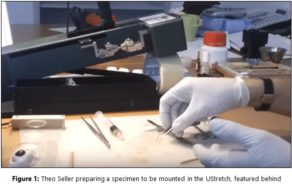
TS: First of all we have the eye of the pig. Then we do the treatment; applying some riboflavin on the top, doing some radiation – whatever treatment is appropriate for the test. When we have done that, we just cut out a piece of the cornea. The width is 3mm and this has already been treated [Figure 1], and if you want to measure it afterwards we just get it into the machine and then we do the stress-strain measurements. We first apply a preload of about 50 millinewtons that simulates the normal eye pressure. Afterwards we do the stress-strain measurement – we want to stress the cornea for twelve percent or we want to apply the strain for twelve percent.
CH: And is that a physiologically relevant strain for the cornea?
TS: It just shows the stiffness. You just want to compare the cornea with and without treatment. We want to see what force we have to apply to get a strain of twelve percent.
CH: Presumably, depending on the nature of the treatments or how much UV exposure and/or how you do the injections of the riboflavin, you get different amounts of stiffness in the cornea?
TS: Exactly, and the UStretch is quite good because it measures really precisely. I think it’s about one millinewton that we can measure. So, we can already detect very little differences between the different treatments; if we use rings in the cornea to get into the riboflavin or we just apply it on the top, we can really achieve quite precise measurements.
CH: That’s very interesting research. Thanks a lot for sharing with us today what you’re doing here at IROC.
Watch the interview online: https://www.youtube.com/watch?v=U3o8efBviSw
Learn more about CellScale’s UniVert.



