
Retinal Tissue Mechanical Stimulation Using a Custom MechanoCulture System

Lund University
Department of Ophthalmology
Project Background
The retina exists in a finely regulated biomechanical environment. Conditions such as retinal detachment, glaucoma, and degenerative diseases involve significant alterations in mechanical load. Despite this, the role of tissue level mechanics in retinal health is not fully understood, in part because of the difficulty associated with applying controlled deformation to such delicate tissue.
A research group at Lund University approached CellScale with the goal of developing custom mechanobiology fixtures capable of:
- In vitro retinal tissue mechanical stimulation
- Applying reproducible retinal stretch testing to isolated retinal segments
- Accommodating extremely fragile tissue during mounting
- Enabling optical imaging during deformation
- Using an existing MechanoCulture B1 test frame as the actuation platform
The challenge was to design a custom mechanobiology fixture that preserved tissue viability and geometry while enabling quantifiable retinal biomechanics studies.

The Challenge
1. Retinal Fragility
Retinal explants are thin, highly fragile, and prone to tearing during handling. Standard mechanobiology gripping fixtures could not be used without risking damage.
2. Need for Radial Stretch in a Controlled Geometry
The research team required a custom attachment that could apply radial or quasi uniform stretch through multiple anchor points without distorting the overall tissue shape.
3. Imaging Compatibility
Simultaneous imaging during stretch was essential for evaluating tissue responses and monitoring deformation. The standard MechanoCulture B1 culture well was not optimized for optical clarity.
4. Integration With an Existing Actuator System
The solution needed to attach directly to the group’s MechanoCulture B1 without modifying the core mechanical components.
Custom Solution Developed by CellScale
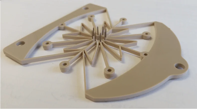
Custom Radial Attachment for Delicate Retinal Mounting
A bespoke specimen holder was designed with:
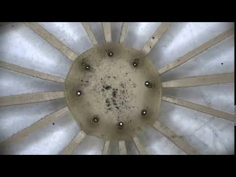
- eight slender anchoring pins arranged in a circular pattern
- flexible supporting arms that minimized stress concentrations
- a low mass structure tailored to prevent tissue distortion
This design allowed researchers to secure porcine retinal tissue reliably while applying controlled retinal stretch testing.
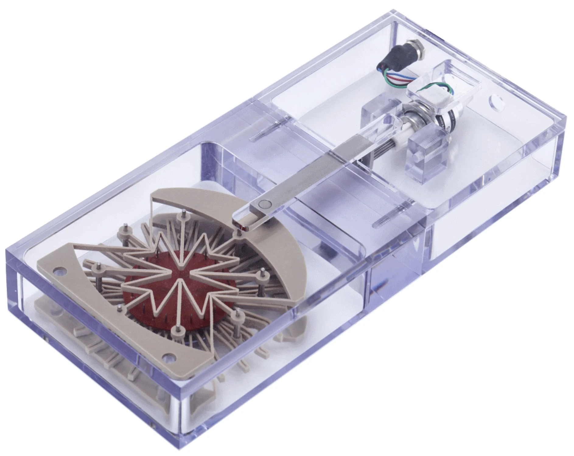
Transparent Polycarbonate Culture Well
The original MCB1 culture well was replaced with a highly polished polycarbonate version, improving optical clarity for both brightfield and fluorescence imaging.
This modification became valuable for other users and was later adopted more broadly outside of retinal biomechanics applications.
Seamless Integration With MechanoCulture B1 Actuation
The custom fixture mounted directly onto the B1 frame, which converted linear actuator motion into radial deformation via the attachment arms.
Iterative Engineering Development
Three iterations of the attachment were produced to optimize:
- the stiffness of the support arms
- anchoring pin spacing
- imaging accessibility
- ease of loading delicate tissue
The final design provided reliable, reproducible retinal tissue mechanical stimulation while maintaining tissue viability throughout multi day culture and stimulation studies.
Hydrated Testing Environment
Testing could be performed directly in media, preserving physiological conditions.
This configuration preserved the MicroTester’s micro-Newton sensitivity while adding new orientation and throughput capabilities tailored to the lab’s workflow.
Results and Impact
The customized MechanoCulture B1 bioreactor system enabled:
1. Precise radial stretch of retinal explants
Researchers were able to apply controlled levels of deformation to study mechanobiological responses to retinal stretch testing.
2. Improved tissue viability during culture
Retinas maintained structural integrity significantly better under controlled stretch compared to unstretched controls, as demonstrated by findings presented at ARVO.
3. Real time imaging during mechanical stimulation
The transparent chamber allowed visualization of retinal structure, deformation and cellular responses during testing.
4. A new platform for retinal biomechanics research
The method supported work presented in an abstract at the 2019 ARVO Annual Meeting, which demonstrated improved retinal survival at physiologically relevant stretch levels.
5. Validation of MechanoCulture systems for custom mechanobiology
Although the MCB1 is no longer produced, this project demonstrates that MechanoCulture systems (TX, T6, TR, and J1) can be adapted to unconventional tissues and experimental needs through custom fixturing.
Key Capabilities Enabled
Controlled retinal tissue mechanical stimulation
Imaging compatible transparent culture environment
Custom fixturing for non standard biological specimens
Reproducible multi point mounting
Seamless integration with MechanoCulture actuators
Related Works
TITLE
Stretch with precision - a novel technique for biomechanical modulation of the retina in vitro
JOURNAL
Investigative Ophthalmology & Visual Science
APPLICATIONS
RESEARCH SUMMARY
This study presented a reproducible in vitro method for applying controlled radial stretch to adult porcine retinal explants, with the goal of improving experimental access to retinal biomechanics. Because the retina exists within a tightly regulated mechanical environment, disruptions in tissue-level forces may contribute to ophthalmological conditions such as retinal detachment and glaucoma. However, the mechanisms linking altered mechanical loading to retinal degeneration remain poorly understood.
To investigate the role of lateral tension in retinal survival, porcine retinal segments were cultured on elastic EPTFE membranes and mounted in a modified CellScale MCB1 biomechanical stimulation system. The system converted axial actuator motion into radial stretch through a deformable scaffold and anchoring-pin arrangement, allowing retinal explants to be held at defined stretch levels ranging from 0% to 16% over a five-day culture period. After culture, specimens were fixed, sectioned, and stained to assess retinal structure and survival of neuronal cell populations.
Unstretched retinal explants showed extensive tissue degradation, including widespread pyknosis and loss of normal laminar architecture. In contrast, stretched retinas demonstrated improved preservation of retinal structure, including maintenance of the inner nuclear layer. Retinas maintained at 4%, 8%, and 16% stretch also showed significantly greater survival of cells with ganglion cell morphology compared with unstretched controls.
These findings suggest that lateral mechanical tension may be an important factor in maintaining retinal tissue integrity in vitro. The study also demonstrated that controlled mechanical stretching can provide a useful experimental platform for retinal biomechanics research, supporting future investigations into how mechanical disturbances contribute to retinal disease and tissue degeneration.
Citation: Jens Nääv Ottosson, Linnea T Taylor, Fredrik K Ghosh; Stretch with precision – a novel technique for biomechanical modulation of the retina in vitro. Invest. Ophthalmol. Vis. Sci. 2019;60(9):2361.
Interested in a Similar Custom Solution?
CellScale engineers design custom mechanobiology fixtures and environments for tissues that cannot be tested using standard mechanical testing equipment.