
[et_pb_section fb_built=”1″ _builder_version=”4.16″ global_colors_info=”{}”][et_pb_row _builder_version=”4.16″ background_size=”initial” background_position=”top_left” background_repeat=”repeat” global_colors_info=”{}”][et_pb_column type=”4_4″ _builder_version=”4.16″ custom_padding=”|||” global_colors_info=”{}” custom_padding__hover=”|||”][et_pb_text _builder_version=”4.16″ global_colors_info=”{}”]In our In the Lab blog series, we catch up with researchers using CellScale systems.
In this installment of the blog, Caleb Horst chats with Shahnaz Javani in the Cardiac Biomechanics Lab of Professor Ali Azadani at the University of Denver. Shahnaz is studying the mechanical properties of heart tissue using the BioTester Planar Biaxial Testing Machine.
Caleb Horst: Welcome, Shahnaz. Thanks for taking the time to speak with me today. I was hoping you could first tell me a little bit more about your research?
Shahnaz Javani: The Cardiac Biomechanics Laboratory is focused on applied and translational research in cardiovascular diseases, so we use modern engineering tools to develop new therapeutic and diagnostic devices. To better understand the physiology and pathophysiology of the heart, and to be able to develop new treatments and therapeutic methods, we need to first know the mechanical properties of the normal heart. So my research specifically is focused on regional mechanical properties of cardiac muscle.
CH: That makes sense, and so you’re obviously doing some mechanical testing as part of your research and you’re using the CellScale BioTester. Can you tell me more about the types of specimens you’re working with and the test protocols you’re using and what types of tissues and so on?
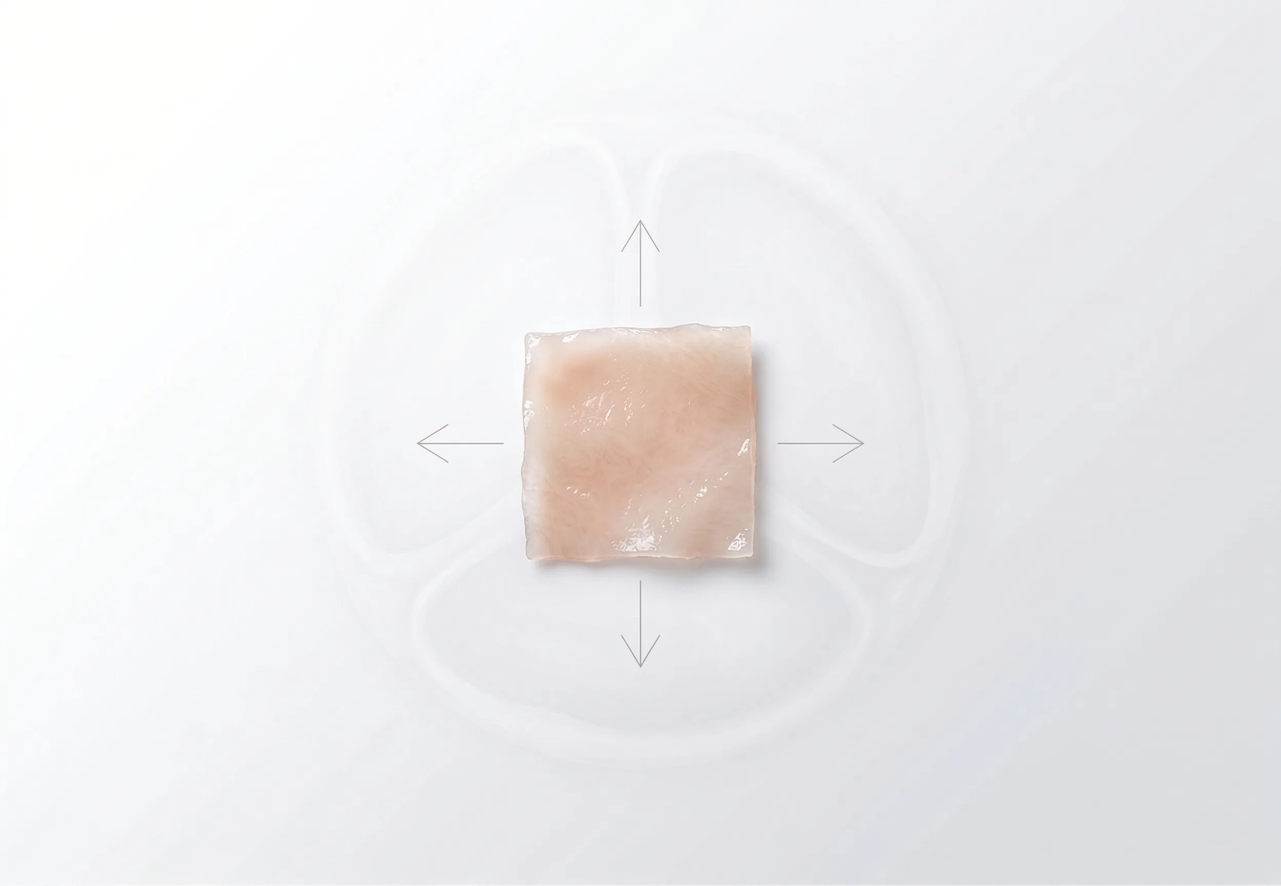
SJ: First of all we know that ovine heart closely resembles human heart from the point of view of physiology and anatomy. So what I do is that I excise square samples from different anatomical regions of the ovine heart, including the ventricles, the atria, and the atrial appendages. The sample dimensions are around 10 by 10 millimeters in plane and two millimeters thick, and they are cut in such a way that two of the edges are parallel to the orientation of the fiber bundles. The samples are then sprinkled with graphite to provide reference points to be easily tracked by the image analysis software. Then, after that, the samples are mounted on the BioTester, where they are immersed into a saline solution of 37 degrees Celsius to simulate the biological environment. After that the samples are subjected to ten cycles of preconditioning followed by one cycle of equibiaxial stretch [Figure 1].
[/et_pb_text][et_pb_image src="https://cellscale.com/wp-content/uploads/2018/10/Blog-6-Fig-1.png" align="center" align_tablet="center" align_phone="center" align_last_edited="on|desktop" _builder_version="4.16" global_colors_info="{}"][/et_pb_image][et_pb_text _builder_version="4.16" global_colors_info="{}"]CH: So once you’ve collected this data then you must have to do some sort of analysis to understand it and get meaningful conclusions. What types of analysis are you doing with the data?
SJ: The stress-strain data obtained from the tests are then fitted to a Fung-type exponential strain energy function and the material constants are obtained [Figure 2]. Strain energy function is a function that relates the strain energy density of a material to the deformation gradient. So having the material constants, the next step would be to compare different regions of the heart based on the tissue stiffness. And of course these material constants will be then used in our finite element models.
[/et_pb_text][et_pb_image src="https://cellscale.com/wp-content/uploads/2018/10/Blog-6-Fig-2.png" align="center" align_tablet="center" align_phone="center" align_last_edited="on|desktop" _builder_version="4.16" global_colors_info="{}"][/et_pb_image][et_pb_text _builder_version="4.16" global_colors_info="{}"]
CH: I see. I also wondered – you probably had a chance to work with a number of different pieces of equipment and types of testing in your lab. Can you tell me how your experiences have been with the BioTester?
SJ: The BioTester is very user-friendly and easy to work with. When I first started working in this lab I spent around an hour learning how to work with it and since then I’ve been using this device to run tests every week. I can easily and rapidly mount the specimens, and I can use it to test a variety of sample sizes. Also, the BioTester software displaying live-time force and displacement plot is a very helpful option, enabling me to monitor the data output in real time.
CH: Great, I’m glad you enjoy those features! I know you haven’t been using it too long, just a number of months, but can you tell me what you’ve learned in that time, what kind of conclusions and what kind of progress you’re making with your research?
SJ: Although I’ve been using this device for only a few months, in this short period of time I’ve been able to collect enough data for my papers to be published in the near future. I was able to compare all the chambers of the heart based on tissue stiffness as well as different portions of each chamber. For example, I found that tissue from a ventricle is significantly stiffer than that from the atria, as well, my results show that atrial appendages are not significantly different from their corresponding atria while they are significantly different from the ventricles.
CH: Thanks so much, Shahnaz. I really do appreciate you taking the time sharing that with me, and by extension, the rest of the world. And I really think you’re doing a great job there. I appreciate it. Once again, thank you.
Watch the interview online: https://www.youtube.com/watch?v=rWLlQaG8Emg
CTA: Learn more about CellScale’s BioTester or check out some of our customers’ publications.
[/et_pb_text][/et_pb_column][/et_pb_row][/et_pb_section]